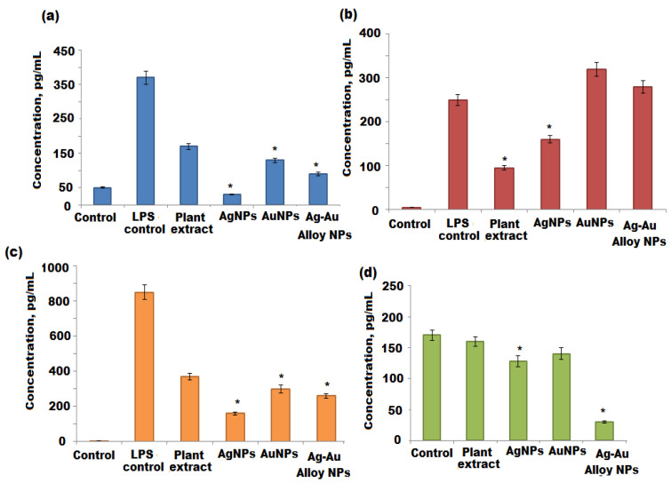
Figure 10.
Quantification of cytokine release from THP1 and NK92 cells following treatment with A. racemosus, Ag, Au, and Ag-Au bimetallic alloy nanoparticles. (a–c) THP1 cells were stimulated with lipopolysaccharide (LPS) for 6 h. The LPS containing medium was then replaced by the respective treatments and the cells were incubated for another 18 h, after which the cytokine production (IL-1β, IL-6, and TNF-α) was quantified by ELISA. (d) NK92 cells were exposed to the respective treatments for 24 h, after which the IFN-γ production was quantified by ELISA. Statistical significance (p < 0.05) compared to the the negative control. * Statistical significance (p < 0.05) compared to the 6 h treatment with LPS (LPS control).

