
Figure 3.
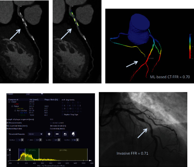
Coronary CT angiography in a 54-year-old man without known coronary artery disease. (a) Automatically generated curved multiplanar reformations showing >50% stenosis of the proximal LAD (arrow). (c) 3-Dimensional color-coded mesh shows a CT-FFR value of 0.70, indicating ischemia of the underlying stenosis (arrow). (b, d) Color-coded automated plaque assessment of the lesion demonstrating the predominantly calcified composition of the atherosclerotic atheroma. (e) Invasive coronary angiography confirms obstructive stenosis of the LAD (arrow) with an FFR of 0.70.