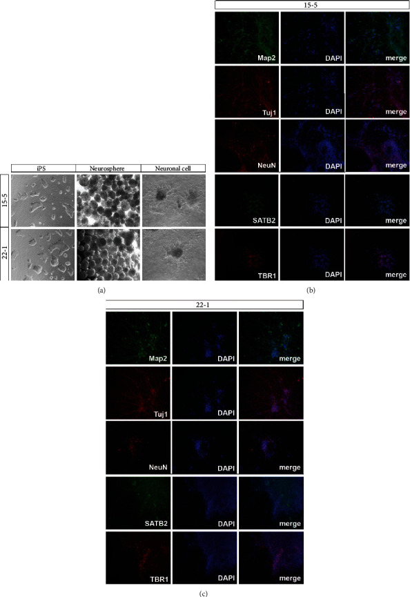
Figure 3.

The result of differentiation. A large number of neurospheres could be observed under the microscope (a). After 28 days of differentiation and cultivation, the neuronal markers Tuj1, Map2, and NeuN and cortical neuronal markers SATB2 and TBR1 were detected by immunofluorescence, which showed that Tuj1, Map2, NeuN, SATB2, and TBR1 were positively expressed (b, c). n = 3.
