



Abstract
The two branches of the Kennedy pathways (CDP-choline and CDP-ethanolamine) are the predominant pathways responsible for the synthesis of the most abundant phospholipids, phosphatidylcholine and phosphatidylethanolamine, respectively, in mammalian membranes. Recently, hereditary diseases associated with single gene mutations in the Kennedy pathways have been identified. Interestingly, genetic diseases within the same pathway vary greatly, ranging from muscular dystrophy to spastic paraplegia to a childhood blinding disorder to bone deformations. Indeed, different point mutations in the same gene (PCYT1; CCTα) result in at least three distinct diseases. In this review, we will summarize and review the genetic diseases associated with mutations in genes of the Kennedy pathway for phospholipid synthesis. These single-gene disorders provide insight, indeed direct genotype-phenotype relationships, into the biological functions of specific enzymes of the Kennedy pathway. We discuss potential mechanisms of how mutations within the same pathway can cause disparate disease.
Keywords: lipid, metabolism, genetic disease, inherited, membrane, phospholipid, phosphatidylcholine, phosphatidylethanolamine
Overview of the Kennedy pathways for phospholipid synthesis
The Kennedy pathways for phospholipid synthesis were first elucidated by Eugene Kennedy and colleagues in the late 1950s and early 1960s and are the major routes by which mammalian cells synthesize the glycerophospholipids phosphatidylcholine (PC) and phosphatidylethanolamine (PE), the two most abundant phospholipids in mammalian cells (1) (Fig. 1). Because high-energy molecules, cytidine diphosphocholine (CDP-choline) and cytidine diphosphoethanolamine (CDP-ethanolamine) are formed as precursors for the synthesis of PC and PE, respectively, the two branches of the Kennedy pathways are often referred to as the CDP-choline and CDP-ethanolamine pathways.
Figure 1.
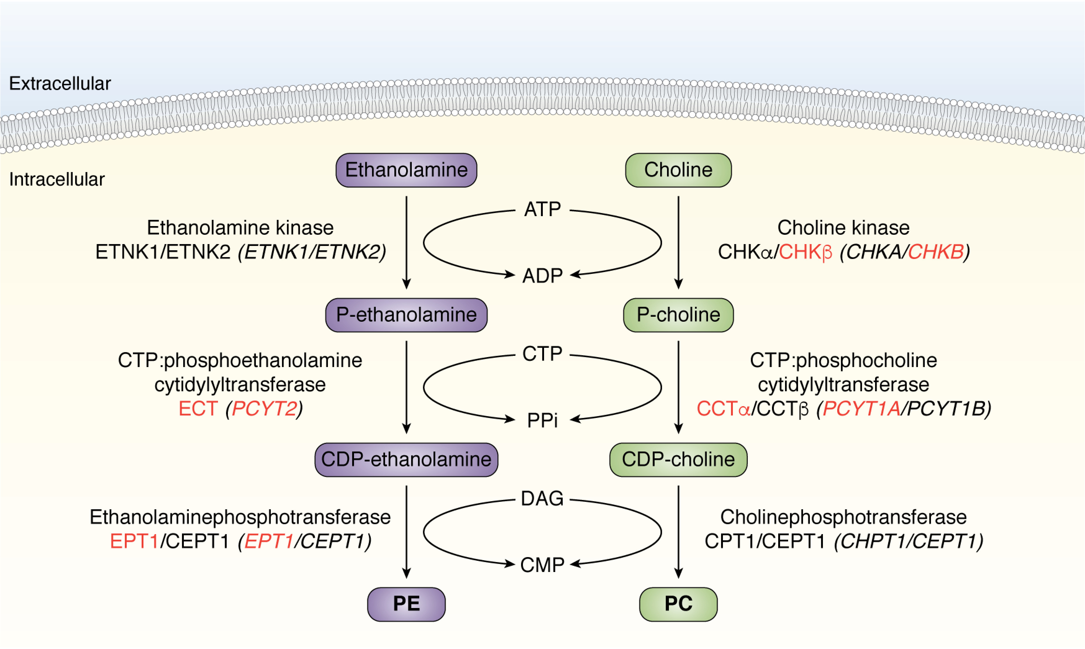
PC and PE biosynthesis via two branches of the Kennedy pathway. The CDP-choline branch for PC synthesis (green) and CDP-ethanolamine branch for PE synthesis (purple). The standardized human names for each protein (Roman type) and gene (italic type) are indicated. Mutations in four genes (red type) have been demonstrated to cause inherited genetic diseases in humans. PC, phosphatidylcholine; PE, phosphatidylethanolamine; DAG, diacylglycerol.
The Kennedy pathways are essential for life at the cellular and organismal levels. The discovery that there are two enzymatic isoforms, encoded by different genes, for almost every Kennedy pathway step was initially puzzling and by in large still is. Some isoforms are ubiquitous and in and of themselves are essential for life, whereas others are not, and this appears to correlate with a more limited tissue/cell type distribution and/or substrate specificity for some isoforms. The recent identification of inherited diseases due to mutations in genes encoding Kennedy pathway enzymes, and the phenotypes associated with each, is beginning to shed light on the specific physiological roles of Kennedy pathway enzymes. To date, of the 10 genes in the two branches of the Kennedy pathway, mutations in four genes have been demonstrated to cause inherited genetic disease in humans—CHKB, PCYT1A, PCYT2, and EPT1 (SELENOI).
PC comprises ∼50% of total mammalian cell phospholipid and is the major lipid building block of membranes. PC also serves as a source of numerous second messenger molecules, such as diacylglycerol, phosphatidic acid, and lysophospholipids (2–4). The first enzymatic step in the CDP-choline branch of the Kennedy pathway is the phosphorylation of choline by choline kinase to produce phosphocholine. In humans, two separate genes, CHKA and CHKB, encoding choline kinase α (CHKα) and choline kinase β (CHKβ), catalyze this reaction. Both of these isoforms are present in the cytoplasm and, according to the human protein atlas, do not appear to have tissue specificity as far as expression of mRNA or presence of protein. The second step, considered to be the rate-limiting reaction for the CDP-choline pathway (5), utilizes phosphocholine and CTP to form CDP-choline with the release of pyrophosphate and is catalyzed by CTP:phosphocholine cytidylyltransferases. Two enzymes encoded by different genes, PCYT1A and PCYT1B encoding CCTα and CCTβ, catalyze this reaction. PCYT1A expression appears to be ubiquitous, whereas PCYT1B expression is enhanced in the brain, in particular the cerebral cortex, and in testis. Both CCT proteins are amphipathic, with the interesting difference that CCTα is primarily internuclear and associates with nuclear membranes, whereas CCTβ is extranuclear and associates with endoplasmic reticulum (ER) membranes. The final step of the CDP-choline pathway is the transfer of phosphocholine from CDP-choline onto a diacylglycerol (DAG) backbone to form PC, and this is catalyzed by cholinephosphotransferases. As per each step of the CDP-choline pathway, two proteins encoded by two separate genes, CHPT1 and CEPT1, which encode CPT1 and CEPT1, can catalyze this reaction (6–8). CHPT1 and CEPT1 expression is ubiquitous. Both are integral membrane-bound proteins with CPT1 localized to the Golgi and CEPT1 to the ER.
PE is the second most abundant phospholipid in most cells, where it comprises 25% of total phospholipid. Beyond its role in maintaining membrane permeability barriers, because of its conical shape (a small polar headgroup relative to the cross-section of the hydrophobic tails), PE can form reverse nonlamellar structures and acts as an intermediate in membrane fusion and fission events via its ability to introduce inverse membrane curvature. The CDP-ethanolamine branch of the Kennedy pathway for PE synthesis parallels those of the CDP-choline pathway for PC synthesis. Ethanolamine is phosphorylated by two cytosolic ethanolamine-specific kinases encoded by ETNK1 and ETNK2 to produce phosphoethanolamine. It is noteworthy that both CHKα and CHKβ also possess ethanolamine kinase activity in vitro, although it is unclear whether CHKα or CHKβ can synthesize PE in vivo (9, 10). ETNK1 and ETNK2 are both cytoplasmic proteins expressed ubiquitously with ETNK2 expression enhanced in the kidney and testis. In the second step, CTP:phosphoethanolamine cytidylyltransferase, encoded solely by the PCYT2 gene, converts phosphoethanolamine and CTP to CDP-ethanolamine and pyrophosphate. This enzyme is soluble and is the only enzyme within the Kennedy pathway that does not have a known second isoform and thus not surprisingly is ubiquitously expressed. The final reaction of the pathway for PE synthesis is the conversion of CDP-ethanolamine and DAG to PE by ethanolaminephosphotransferases, of which there are two isoforms encoded by EPT1 (also known as SELENOI) and CEPT1 (6–8). Both are ubiquitously expressed and are integral membrane-bound proteins. EPT1 synthesizes PE exclusively and is localized to the Golgi, and as mentioned earlier in this review, CEPT1 can also synthesize PC and is localized to the ER.
Interestingly, although there is clear tissue-specific distribution of some isoforms for enzymes within the Kennedy pathways, differences in tissue distribution do not appear to be the major driver behind the inherited diseases of the Kennedy pathway. It is not known whether there is metabolic channeling of substrates along any specific pathway (e.g. CHKα/CCTα/CEPT1) that could explain the observed Kennedy pathway inherited diseases. Similarly, differences in subcellular localization of isoforms that can catalyze the same enzymatic reaction do not appear to be the cause of inherited diseases of the Kennedy pathway, although this has yet to be completely ruled out.
Mutations in the first step of the CDP-choline pathway for PC synthesis cause an inherited muscular dystrophy
Mutations in the CHKB gene (OMIM 612395) cause a progressive megaconial congenital muscular dystrophy. CHKB-mediated muscular dystrophy is an autosomal recessive disorder caused by loss-of-function mutations (phenotype OMIM number 602541). Of the 30 genes known to cause muscular dystrophy, this is the only form that is due to a defect in membrane synthesis. CHKB-mediated muscular dystrophy presents as a rostral-to-caudal gradient and is often accompanied by neonatal hypotonia, global developmental delays without brain malformation, ichthyosis-like changes of skin, and dilated cardiomyopathy. In affected muscle, there are mitochondrial structural changes including enlarged mitochondria at the periphery of muscle fibers and loss of mitochondria at the center (8, 11–18).
Mutations in the CHKB gene that cause muscular dystrophy have been found in 10 of the 11 exons of the CHKB gene (17) (Fig. 2). Functional analysis of these mutations shows that they result in choline kinase activity that was either undetectable or severely decreased (11, 13, 19, 20). Interestingly, studies of cells from CHKB patients and muscle from Chkb−/− knockout mice determined that there was only a very minor decrease in PC content, and this decrease was also seen in unaffected muscle (11). In addition, in CHKB patients and Chkb−/− knockout mice, not all tissues are affected (18), suggesting that residual choline kinase activity encoded by CHKA may rescue these unaffected cell types. Consistent with this hypothesis, adenovirus transfection of either Chkb or Chka into Chkb−/− knockout mice rescued the muscular dystrophy (21), indicating that increasing the dose of the Chka isoform in mice is able to protect against loss of function of CHKB. It has yet to be determined whether a difference in CHKA expression is the cause of the rostral-to-caudal gradient in CHKB-mediated muscular dystrophy, although it is reasonable to assume that this could be the case. If so, determining why CHKA expression is differentially regulated in different tissues, or even in muscles in different regions of the body, could lead to a better understanding of CHKB-mediated muscular dystrophy and point to a potential therapeutic route.
Figure 2.
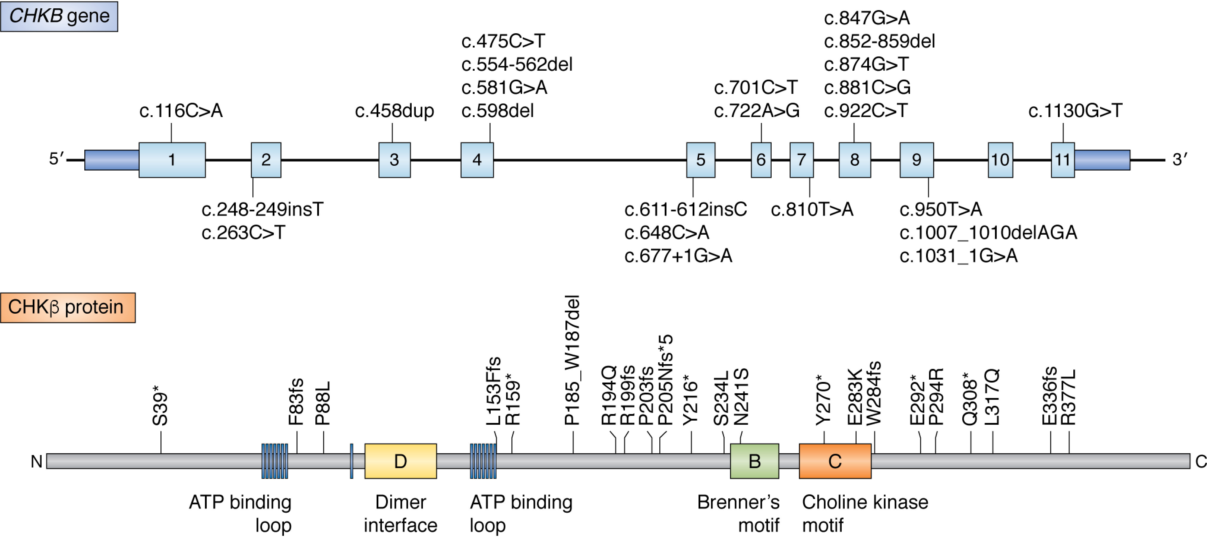
Summary of the currently reported mutations in human CHKB. Top, schematic drawing of the CHKB gene. Dark blue rectangles, exon noncoding regions; light blue rectangles, exon coding regions. Bottom, amino acid sequence of the CHKB (CHKβ) protein. The boxes highlight amino acids forming the ATP-binding loop (blue) and dimer interface (yellow). The conserved phosphotransferase Brenner's motif (green) and choline kinase motif (orange) are also indicated. Mutations in the CHKB gene cause a progressive megaconial congenital muscular dystrophy.
Regardless of the expression of CHKA in affected muscle in CHKB-mediated muscular dystrophy, one interesting observation that needs to be reconciled is that the level of PC does not appreciably change in muscle cells lacking CHKB; nor is there a difference in PC level between affected and unaffected muscle along the rostral-to-caudal gradient of this form of muscular dystrophy (18). PC can be taken up by cells from extracellular sources, which could explain why there is no appreciable change in PC mass. Indeed, the most notable change in phospholipid mass in muscle from both Chkb−/− knockout mice (22) and humans (11) to date is a slight increase in the level of PE, with the altered PC/PE ratio being proposed to affect membrane function in muscle due to the different biophysical characteristics of each phospholipid (11, 22). A second explanation that has yet to be tested is how inactivation of CHKB affects metabolites of the CDP-choline pathway itself. The two most obvious changes would be an in increase in choline, the substrate of the choline kinase reaction, as well as an inability to consume CDP-choline metabolites by enzymes downstream of the choline kinase step. The most notable of these downstream metabolites would be the consumption of DAG by the terminal CPT1/CEPT1 cholinephosphotransferase step (Fig. 1). Increased DAG has a plethora of effects on lipid metabolism (and signaling), including its conversion to triglyceride, which would result in an accumulation of fatty lipid droplets in affected muscle. A more thorough investigation of the effect of CHKB inactivation in affected versus unaffected muscle on global lipid metabolism should aid in determining the mechanism by which a defect in PC synthesis can cause muscular dystrophy.
Mutations in the rate-determining step for PC synthesis can cause three different inherited diseases
PCYT1A (OMIM 123695) encodes the enzyme CTP:phosphocholine cytidylyltransferase A (CCTα) responsible for converting phosphocholine into CDP-choline in the Kennedy pathway for PC synthesis. Recently, three distinct autosomal recessive diseases appear to be caused by mutations in the PCYT1A gene, spondylometaphyseal dysplasia with cone-rod dystrophy (SMD-CRD) (phenotype OMIM number 608940), Leber congenital amaurosis (LCA), and congenital lipodystrophy with severe fatty liver disease (CL-FLD) (23–26) (Fig. 3A). How mutations in the same gene, PCYT1A, can cause three different inherited diseases is an interesting biochemical and biological problem.
Figure 3.
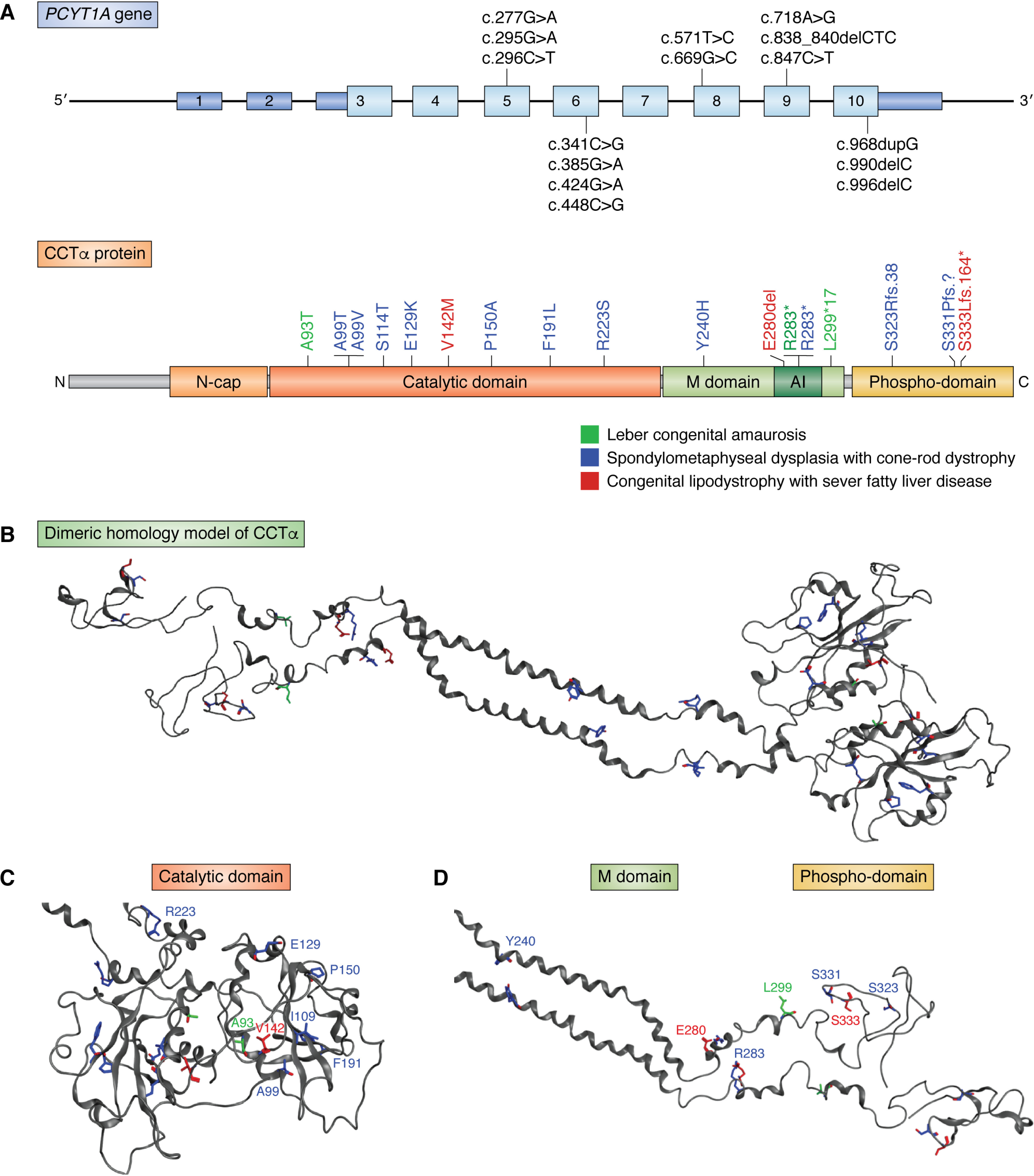
Summary of the currently reported mutations in human PCYT1A. A, top, schematic drawing of the PCYT1A gene. Dark blue rectangles, exon noncoding regions; light blue rectangles, exon coding regions. Bottom, amino acid sequence of the CCTα protein. The boxes highlight domain structure of CCTα. The N-cap (light red) is common to all CCTs and contains the nuclear localization signal. Red, catalytic domain. Green, membrane-binding domain (M domain), which contains the autoinhibitory helix (AI). Orange, phosphorylation region that is the site of up to 16 phosphoserines (Phospho-Domain). The sites of all disease-linked mutations are depicted. The various PCYT1A phenotypes corresponding to the mutations are highlighted: spondylometaphyseal dysplasia with cone-rod dystrophy (dark blue), congenital lipodystrophy and severe fatty liver disease (red), and Leber congenital amaurosis (green). B, three-dimensional model of human CCTα with known inherited disease-causing mutations noted. A dimeric homology model of residues 40–367 of CCTα was built in the Molecular Operating Environment software suite (version 2019.0102) using the following Protein Data Bank structures: 4MVC, 4MVD, 3FDE, 5H53, and 4WJ4. A short molecular dynamics simulation of 1 ns was used to relax the system. C, homology model of the catalytic domain of CCTα with amino acid mutation site locations identified. D, homology model of the membrane-binding and C-terminal phosphodomain of CCTα with inherited disease amino acid mutation locations highlighted.
Mutations in PCYT1A that can cause SMD-CRD consist of single amino acid changes (A99T, A99V, S114T, E129K, P150A, F191L, R223S, and Y240H), a nonsense mutation (R283*), and two frameshift mutations (S323Rfs.38 and S331Pfs?) (27). The disease is autosomal recessive with some patients possessing homozygous mutations and some patients possessing biallelic heterozygous variants. SMD-CRD is characterized by severely short stature, progressive bowing of the lower limbs, flattened vertebral bodies (platyspondyly), metaphyseal abnormalities, and visual impairment caused by cone and rod dystrophy (26). There are 10 described SMD disorders, with only two of those affecting vision with one of these being PCYT1A-mediated SMD-CRD (23, 28).
Mutations in PCYT1A believed to cause LCA consist of a single amino acid change (A93T), a nonsense mutation (R283*, also observed in SMD-CRD patients), and a mutation (L299*17) that inserts 17 foreign amino acid residues at the C terminus of CCTα (27). PCYT1A-mediated LCA demonstrates an autosomal recessive inheritance pattern with known mutations being heterozygous biallelic. LCA is a severe retinal dystrophy causing severe visual impairment or blindness before the age of one (29). LCA is characterized by nystagmus, poor pupillary light response, and retinal function that is undetectable or severely abnormal (30). To date, mutations in more than 20 genes have been found to be associated with LCA.
PCYT1A mutations that cause CL-FLD consist of a single amino acid deletion (E280del), a single nucleotide change (V142M), and a frameshift mutation (S333Lfs.164) (27). This is also an autosomal recessive disease with heterozygous biallelic inheritance. This disease is characterized by the childhood presentation of lipodystrophy (abnormal distribution of fat), severe nonalcoholic fatty liver disease, type II diabetes, dyslipidemia (mainly very low high-density lipoprotein level), and modestly short stature (24).
Of the Kennedy pathway enzymes whose mutation can cause an inherited disease, the structure has only been solved for mammalian (rat) CCTα. Previous work used computational modeling to map each disease-causing amino acid variant from its linear two-dimensional representation onto a three-dimensional structure (27). Here, we used a different computational method to predict the human CCTα structure based on rat CCTα (Fig. 3, B–D) and pinpoint the location of each amino acid residue whose mutation causes a specific inherited disease. Both methods noted that disease-causing mutations in CCTα did not correlate with a specific region in the tertiary structure of CCTα. How these changes could cause inherited disease may be gleaned from specific knowledge of CCTα domains and their structure and function.
Regarding CCTα structure and function, CCTα has four distinct domains: 1) an N-terminal domain containing the nuclear localizing signal; 2) a catalytic domain; 3) a membrane-binding domain; and 4) an unstructured tail with multiple residues that undergo phosphorylation (31, 32). The membrane-binding domain contains an autoinhibitory segment that interacts with the catalytic domain to inhibit activity when the enzyme is not bound to a membrane (27, 33). The mutations in the alleles of PCYT1A that can cause each inherited disease are localized throughout the catalytic and regulatory domains of the encoded CCTα protein (Fig. 3, A–D). In cells, the membrane-bound catalytically active form of CCTα localizes mainly to the nuclear envelope/ER when active and localizes to the nucleoplasm when inactive (34). There is also evidence in some cell types that CCT localizes to the cytoplasm when inactive (35). CCTα homodimerizes (36) with each monomer, contributing amino acid residues to form the active site (Fig. 3, B and C). When the enzyme is inactive, the amphipathic helix of each monomer forms a complex of four helices with two helices of the catalytic domain (27). This complex changes the conformational shape of the enzyme and prevents access to essential catalytic residues in the catalytic core. When the enzyme binds to a membrane, the helices of the membrane-binding domain become embedded in the membrane surface and displace an autoinhibitory domain, enabling substrates to access the catalytic core (27). This process is selective for membranes that have a high negative surface change and loose packing, ensuring that the enzyme is only catalytically active when the cell requires membrane synthesis (37).
Interestingly, studies of Pcyt1a−/− mice demonstrated that total absence of Pcyt1a results in embryonic death as zygotes did not develop past day 3.5 (38). This implies that the human alleles of PCYT1A that cause inherited disease likely do not result in total loss of function. Analysis of the expression of each allele from in patient-derived fibroblast cell lines is incomplete; however, of those analyzed, a decrease in CCTα protein from as little as 15% to as high as 85% was noted. It is known that in fibroblasts, CCTα levels can be reduced by as much as 95% before a decrease in the rate of PC synthesis is observed (39).
A comprehensive analysis of how disease-causing mutations could alter CCTα activity was recently determined using purified protein. Enzyme activity was assessed in the presence and absence of activating lipids to determine basal (soluble) and membrane-activated levels of CCTα activity. The 13 CCTα disease alleles analyzed fell into four classes with different biochemical features. The first group of mutants (V142M and P150A) manifested low activity in cell lysates associated with reduced solubility caused by aberrant folding and aggregation. The second group of mutants (S114T, F191L, R283*, and S333L.fs) had severely reduced expression levels caused by aggregation and potential degradation of misfolded protein. The third group of mutants (A93T, A99T, A99V, and E129K) had moderate solubility but reduced enzymatic activity and thermal destabilization. The fourth group of the mutants (R223S) had low enzymatic activity without fold destabilization. Mutants in the M domain (Y240H and E280del) were considered outliers, with Y240H showing reduced solubility with no functional disability and E280del having elevated constitutive activity and enhanced membrane binding but decreased thermal stability and lower activity in cell lysates. Interestingly, there was no clear effect on CCTα enzyme activity that correlated with mutations that cause a specific inherited disease due to mutations in PCYT1A. As CCTα homodimerizes, and many of the diseases are heterozygous biallelic, further work could include assessing how the biallelic PCYT1A disease-causing PCYT1A mutations, when in combination, affect CCTα activity.
One must also consider the nonenzymatic observations and/or functions that have been described for CCTα as potential contributors to the inherited disease phenotypes. In most cell types, CCTα translocates into the nucleus, whereas in some cells, CCTα is primarily extranuclear. However, the rate of PC synthesis does not appear to be affected by CCTα localization (40, 41). The role of the duality for CCTα localization and its impact on function is not clear and requires further understanding. Within the nucleus, CCTα was determined to regulate the formation of the nucleoplasmic reticulum (NR). The tubules of the NR contain components of the endoplasmic reticulum, nuclear envelope, and cytoplasm. Interestingly, both catalytically dead and constitutively active forms of CCTα result in NR proliferation, indicating that enzyme activity is not essential for expansion of the NR by CCTα (42, 43). The NR expands the total surface area of the nuclear membrane and is purported to increase transport of both proteins and ions into and out of the nucleus. Determining how CCTα regulates NR function may enable an understanding of how PCYT1A mutations, many of which do not appear to affect PC synthesis, could result in an inherited disease.
Mutations in PCYT2 and EPT1 for PE synthesis cause inherited spastic paraplegias
Complex hereditary spastic paraplegia (HSP) (phenotype OMIM numbers 618770 and 618768) can be caused by mutations in PCYT2 (OMIM 602679) coding for CTP:phosphoethanolamine cytidylyltransferase and EPT1 (SELENOI) (OMIM 607915) coding for ethanolaminephosphotransferase, the second and final steps in the CDP-ethanolamine pathway for PE synthesis, respectively. HSP is a group of degenerative, neurological disorders that primarily affect the upper motor neurons in the brain and spinal cord. There are more than 80 different genetic types of HSP, but they all share the common symptom of difficulty walking due to muscle weakness and muscle tightness (spasticity) in the legs; the term complex HSP is used when these features are associated with other neurological or nonneurological features, such as peripheral nerve impairment, muscle atrophy, or intellectual impairment. For PCYT2 and EPT1 forms of complex HSP, these were often accompanied by epileptic seizures (44).
To date, PCYT2 variants that cause HSP (45) are a homozygous nonsense variant c.1129C>T (R377*) and compound heterozygous variants resulting in H244Y and P307L amino acid changes (45) (Fig. 4). The amino acid variants are predicted to result in substitution of highly conserved residues in the catalytic cytidylyltransferase domain of PCYT2 and are predicted to be damaging by multiple in silico tools (27). In patient-derived fibroblasts, the variants studied to date presented with significantly decreased enzymatic activity (45). Consistent with only one homologue of PCYT2 present in humans, studies of a pcyt2 zebrafish knockout (45) and the Pcyt2−/− mouse (46) indicate that complete loss of CTP:phosphoethanolamine cytidylyltransferase function is incompatible with life in vertebrates. This implies that some PCYT2 enzyme activity is likely present in HSP patients with PCYT2 mutations, although this awaits a more thorough analysis.
Figure 4.
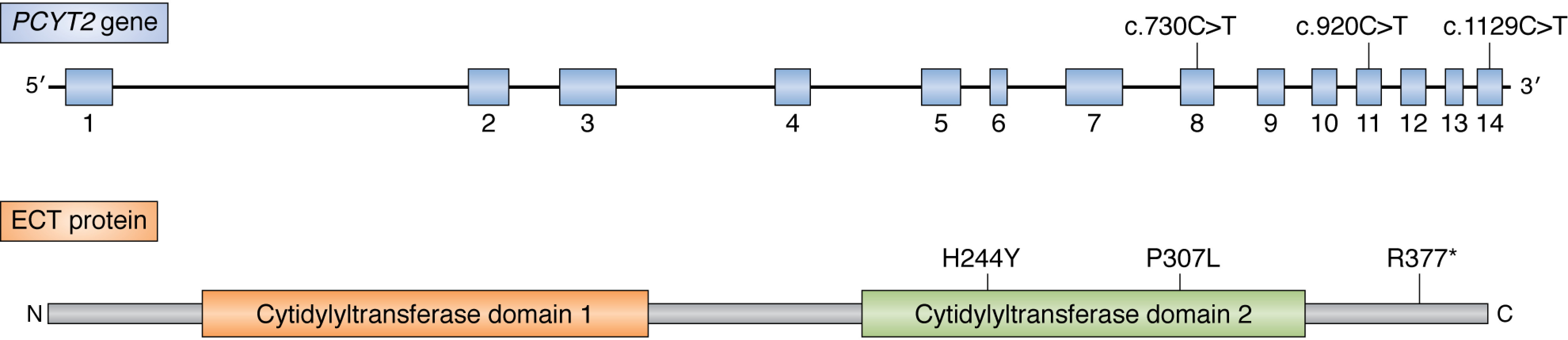
Summary of the currently reported mutations in human PCYT2. Top, schematic drawing of the PCYT2 gene. Exons are shown in light blue. Bottom, amino acid sequence of the ECT protein. The cytidylyltransferase domains I and II are highlighted. Mutations in PCYT2 for PE synthesis cause inherited spastic paraplegias.
In PCYT2 patient fibroblasts, lipidomic analysis revealed a modest reduction in the level of PE to 70% that of controls (45). A reduction in some PE species with unsaturated fatty acids was also observed, as was the level of vinyl-ether–linked plasmenyl-PE (1-alkenyl-2-acyl glycerophosphoethanolamine). Interestingly, there was a large accumulation of ether-linked plasmanyl-PC (1-alkyl-2-acyl glycerophosphocholine) (45).
Mutations have also been identified in EPT1 that cause complex HSP. Thus far, these mutations are autosomal recessive with homozygous mutations present on each allele (47, 48) (Fig. 5). Identified mutations in EPT1 that cause HSP are c.732-2A>G, which resulted in splice site defects with premature terminations, and a point mutant that results in an R112P amino acid change (47, 48). The R112P mutation resides within the CDP-alcohol phosphotransferase consensus sequence, a catalytic motif found in numerous lipid biosynthetic enzymes that is DX2DGX2ARX8GX3DX3D, where X is any amino acid that is required for catalytic activity by this class of enzymes, and analysis of enzyme activity was consistent with an almost complete loss of function (8). A deleterious effect of the exon-skipping mutation on EPT1 catalytic activity was observed when analyzed using patient skin fibroblasts (48). Loss of function of EPT1 may be able to be compensated for, in part, by the presence of CEPT1, although this remains to be established. To this end, knockdown of EPT1 or CEPT1 in HEK293 cells resulted in viable cells (49), with metabolic labeling analyses revealing that EPT1 preferentially synthesized plasmenyl-PE and PE species with long-chain unsaturated fatty acids, whereas CEPT1 preferentially synthesized diacyl-PE with shorter and more saturated fatty acids (49). Similarly, analysis of cells derived from EPT1 HSP patients determined that the total level of PE was similar to controls; however, PE species with polyunsaturated fatty acids were reduced (47, 48). Lipidomic analysis of patient-derived skin fibroblasts and EPT1-KO HeLa cells determined that plasmenyl-PE was decreased to half that of controls. Interestingly, plasmanyl-PC species was increased to twice the amount of controls (48). These changes in PE and PC species are similar to those observed in PCYT2-derived patient fibroblasts.
Figure 5.
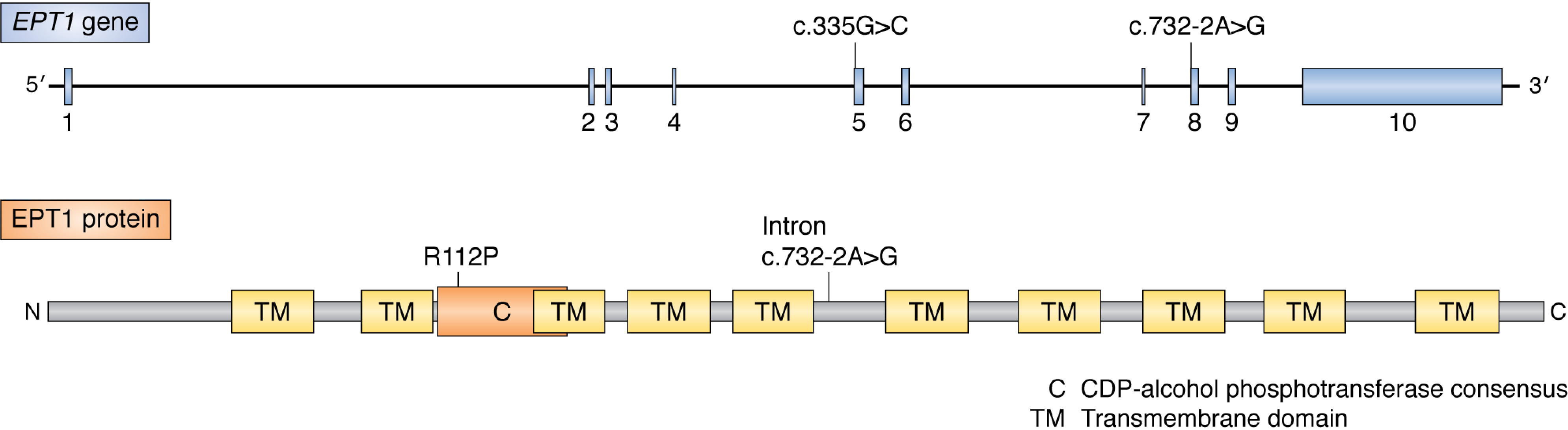
Summary of the currently reported mutations in human EPT1. Top, schematic drawing of the EPT1 gene. Exons are shown in light blue. Bottom, amino acid sequence of the EPT1 protein. The C domain (catalytic motif of the CDP-aminoalcohol phosphotransferase domain) and predicted transmembrane domains (TM) are highlighted. Mutations in EPT1 for PE synthesis cause inherited spastic paraplegias.
Interestingly, EPT1 is a selenoprotein. These are a small number of proteins that contain the 21st amino acid, selenocysteine. Selenocysteine resembles cysteine except that selenium is substituted for sulfur. Another notable selenoprotein is GPX4, a GSH peroxidase that detoxifies unsaturated fatty acids after their damage by reactive oxygen species. Interestingly, mutations in the human GPX4 gene (OMIM 138322) cause a spondylometaphyseal dysplasia (SMD; OMIM 250220) that includes differential neuronal system malformations. Most patients die within a few days of birth. Interestingly, a form of SMD has also been observed in patients with specific PCYT1A mutations. Transgenic mice where the codon encoding the lone selenocysteine in Gpx4 was replaced with a cysteine codon (Gpx4cys/cys) resulted in viable mice; however, they had serious epileptic seizures. Interestingly, the survival of a specific type of interneuron in the Gpx4cys/cys mice was compromised in these cells, and they were highly sensitive to ferroptosis. It was proposed from this study that selenocysteine within a protein confers resistance to overoxidation due to excessive lipid peroxidation. Interestingly, the synthesis of ether-linked lipids and lipids with increased degrees of unsaturation promotes ferroptosis. EPT1 has been demonstrated to preferentially synthesize PE species with ether linkages for their fatty acids and PE with a high level of unsaturated fatty acids. As EPT1 is a selonoprotein and also has been shown to directly synthesize the forms of PE that enable ferroptosis, this opens up the interesting hypothesis that loss of function of EPT1 results in cell death due to an inability to regulate ferroptosis.
The neuronal phenotype associated with decrease/loss of function of PCYT2/EPT1 could also be due, in part, to the increased requirement for PE/plasmenyl-PE in the brain. PE is enriched in the human brain, where it accounts for 45% of the total phospholipid (50, 51), with plasmenyl-PE accounting for 70% of PE in the myelin sheath (52). Plasmenyl-PE protects cells against the damaging effects of reactive oxygen species, and decreasing plasmenyl-PE level could result in cells less able to survive any insults that increase reactive oxygen level (53). Plasmenyl-PE is also a major constituent in membranes that undergo rapid membrane fusion, such as synaptic vesicles. PE, and especially plasmenyl-PE, promote membrane fusion events preferentially over other lipids due to their enhanced ability to form inverted hexagonal phases, with decreased plasmenyl-PE level potentially decreasing synaptic vesicle fusion events (53, 54).
Discussion
Single-gene inherited diseases provide a direct causal genotype-phenotype relationship for the encoded gene. The hereditary diseases associated with single gene mutations in the Kennedy pathways for phospholipid biosynthesis imply specialized roles in muscle, retina, bone, liver, and adipose tissue.
Phospholipid metabolism is highly interconnected with that of other lipids within a cell. In addition, lipids and lipid precursors can be imported into the cell from lipoproteins, BSA, or direct transporters (55). As an example, fatty acids derived from plasma have divergent fates, depending on the metabolic status of the cell. The major metabolic fates of fatty acids are (i) mitochondrial β oxidation to provide energy; (ii) conversion into phospholipids/sphingolipids, where they serve as important structural components of membranes and act as reservoirs for numerous signaling molecules; or (iii) synthesis of neutral lipid species, such as triacylglycerides, and storage in discrete lipid droplets (56). A set of complex regulatory mechanisms has evolved to modify the activity of different enzymes in lipid biosynthesis. These regulatory mechanisms can broadly affect flux into or through a synthetic pathway or specifically affect levels of an individual type of lipid (57). Therefore, mutation in a gene involved in lipid biosynthesis can result in generation of adaptive and maladaptive lipid metabolism (58, 59). An example of these regulatory mechanisms can be found in studies showing that PC level alters activation of SREBP1c, a transcription factor regulating expression of all of the key enzymes involved in fatty acid synthesis. PCYT1A deletion in human hepatocytes reduces PC levels and increases lipogenesis and lipid droplet triacylglyceride accumulation via SREBP1c activation (60). A more thorough lipidomics analysis of Kennedy pathway diseases as they progress over time should point to major metabolic disturbances in lipid metabolism, beyond the Kennedy pathways themselves, that could be major contributors/causes of the disease phenotypes observed.
The impact of nutrition could also alter disease progression. The Kennedy pathway contributes ∼70% of PC produced when choline supply is adequate, with the remaining ∼30% produced in the liver through the PE methylation pathway. However, when choline is limiting in the diet, the PE methylation pathway is crucial for maintaining the supply of PC in the liver, and its subsequent delivery to other tissues via lipoproteins and choline deficiency in mice results in nonalcoholic fatty liver disease (61). The mutations in various genes in the Kennedy pathway might well make the patients more susceptible to the level of choline, ethanolamine, or other lipid precursors in their diet.
Tissue-specific expression of a specific Kennedy pathway gene/protein could point to specific disease phenotypes; however, many of the genes mutated are ubiquitously expressed. Interestingly, almost every step in each Kennedy pathway has two orthologs that can catalyze the same reaction. Increased expression of the unaffected ortholog may protect some cell types from decreased/lost/altered function of the other. For instance, although targeted deletion of the Pcyt1a gene in mice causes early embryonic lethality, Pcyt1a heterozygous mice are viable, with reduction of the Pcyt1a transcript accompanied by up-regulation of Pcyt1b and Pempt transcripts, two other enzymes capable of producing PC (38). Future studies aimed at characterizing the expression pattern and enzymatic activity of different mutations in a tissue-specific manner and/or by generating tissue-specific knockouts will enable insight into the physiological roles of each Kennedy pathway enzyme.
Mutations in individual genes of the CDP-choline and CDP-ethanolamine branches of the Kennedy pathway cause multiple and disparate inherited genetic diseases in humans. Further research will enable more precise genotype-phenotype relationships and enable the determination of pathogenic mechanisms.
Funding and additional information—Work on this topic was supported by the Canadian Institutes of Health Research.
Conflict of interest—The authors declare that they have no conflicts of interest with the contents of this article.
- PC
- phosphatidylcholine
- PE
- phosphatidylethanolamine
- ER
- endoplasmic reticulum
- DAG
- diacylglycerol
- SMD
- spondylometaphyseal dysplasia
- SMD-CRD
- SMD with cone-rod dystrophy
- LCA
- Leber congenital amaurosis
- CL-FLD
- congenital lipodystrophy with severe fatty liver disease
- NR
- nucleoplasmic reticulum
- HSP
- hereditary spastic paraplegia.
References
- 1. McMaster C. R. (2018) From yeast to humans - roles of the Kennedy pathway for phosphatidylcholine synthesis. FEBS Lett. 592, 1256–1272 10.1002/1873-3468.12919 [DOI] [PubMed] [Google Scholar]
- 2. Fagone P., and Jackowski S. (2013) Phosphatidylcholine and the CDP-choline cycle. Biochim. Biophys. Acta 1831, 523–532 10.1016/j.bbalip.2012.09.009 [DOI] [PMC free article] [PubMed] [Google Scholar]
- 3. Exton J. H. (1994) Phosphatidylcholine breakdown and signal transduction. Biochim. Biophys. Acta 1212, 26–42 10.1016/0005-2760(94)90186-4 [DOI] [PubMed] [Google Scholar]
- 4. Ridgway N. D. (2013) The role of phosphatidylcholine and choline metabolites to cell proliferation and survival. Crit. Rev. Biochem. Mol. Biol. 48, 20–38 10.3109/10409238.2012.735643 [DOI] [PubMed] [Google Scholar]
- 5. Sundler R., and Akesson B. (1975) Biosynthesis of phosphatidylethanolamines and phosphatidylcholines from ethanolamine and choline in rat liver. Biochem. J. 146, 309–315 10.1042/bj1460309 [DOI] [PMC free article] [PubMed] [Google Scholar]
- 6. Henneberry A. L., Wistow G., and McMaster C. R. (2000) Cloning, genomic organization, and characterization of a human cholinephosphotransferase. J. Biol. Chem. 275, 29808–29815 10.1074/jbc.M005786200 [DOI] [PubMed] [Google Scholar]
- 7. Henneberry A. L., and McMaster C. R. (1999) Cloning and expression of a human choline/ethanolaminephosphotransferase: synthesis of phosphatidylcholine and phosphatidylethanolamine. Biochem. J. 339, 291–298 10.1042/0264-6021:3390291 [DOI] [PMC free article] [PubMed] [Google Scholar]
- 8. Henneberry A. L., Wright M. M., and McMaster C. R. (2002) The major sites of cellular phospholipid synthesis and molecular determinants of Fatty Acid and lipid head group specificity. Mol. Biol. Cell 13, 3148–3161 10.1091/mbc.01-11-0540 [DOI] [PMC free article] [PubMed] [Google Scholar]
- 9. Gallego-Ortega D., Ramirez de Molina A., Ramos M. A., Valdes-Mora F., Barderas M. G., Sarmentero-Estrada J., and Lacal J. C. (2009) Differential role of human choline kinase α and β enzymes in lipid metabolism: implications in cancer onset and treatment. PLoS ONE 4, e7819 10.1371/journal.pone.0007819 [DOI] [PMC free article] [PubMed] [Google Scholar]
- 10. Chang C. C., Few L. L., Konrad M., and See Too W. C. (2016) Phosphorylation of human choline kinase β by protein kinase A: its impact on activity and inhibition. PLoS ONE 11, e0154702 10.1371/journal.pone.0154702 [DOI] [PMC free article] [PubMed] [Google Scholar]
- 11. Mitsuhashi S., Ohkuma A., Talim B., Karahashi M., Koumura T., Aoyama C., Kurihara M., Quinlivan R., Sewry C., Mitsuhashi H., Goto K., Koksal B., Kale G., Ikeda K., Taguchi R., et al. (2011) A congenital muscular dystrophy with mitochondrial structural abnormalities caused by defective de novo phosphatidylcholine biosynthesis. Am. J. Hum. Genet. 88, 845–851 10.1016/j.ajhg.2011.05.010 [DOI] [PMC free article] [PubMed] [Google Scholar]
- 12. Gutierrez Rios P., Kalra A. A., Wilson J. D., Tanji K., Akman H. O., Gómez E. A., Schon E. A., and DiMauro S. (2012) Congenital megaconial myopathy due to a novel defect in the choline kinase beta gene. Arch. Neurol. 69, 657–661 10.1001/archneurol.2011.2333 [DOI] [PMC free article] [PubMed] [Google Scholar]
- 13. Quinlivan R., Mitsuahashi S., Sewry C., Cirak S., Aoyama C., Mooore D., Abbs S., Robb S., Newton T., Moss C., Birchall D., Sugimoto H., Bushby K., Guglieri M., Muntoni F., et al. (2013) Muscular dystrophy with large mitochondria associated with mutations in the CHKB gene in three British patients: extending the clinical and pathological phenotype. Neuromuscul. Disord. 23, 549–556 10.1016/j.nmd.2013.04.002 [DOI] [PubMed] [Google Scholar]
- 14. Castro-Gago M., Dacruz-Alvarez D., Pintos-Martínez E., Beiras-Iglesias A., Delmiro A., Arenas J., Martín M. Á., and Martínez-Azorín F. (2014) Exome sequencing identifies a CHKB mutation in Spanish patient with megaconial congenital muscular dystrophy and mtDNA depletion. Eur. J. Paediatr. Neurol. 18, 796–800 10.1016/j.ejpn.2014.06.005 [DOI] [PubMed] [Google Scholar]
- 15. Cabrera-Serrano M., Junckerstorff R. C., Atkinson V., Sivadorai P., Allcock R. J., Lamont P., and Laing N. G. (2015) Novel CHKB mutation expands the megaconial muscular dystrophy phenotype. Muscle Nerve 51, 140–143 10.1002/mus.24446 [DOI] [PubMed] [Google Scholar]
- 16. Oliveira J., Negrão L., Fineza I., Taipa R., Melo-Pires M., Fortuna A. M., Gonçalves A. R., Froufe H., Egas C., Santos R., and Sousa M. (2015) New splicing mutation in the choline kinase beta (CHKB) gene causing a muscular dystrophy detected by whole-exome sequencing. J. Hum. Genet. 60, 305–312 10.1038/jhg.2015.20 [DOI] [PubMed] [Google Scholar]
- 17. Chan S. H., Ho R. S., Khong P. L., Chung B. H., Tsang M. H., Yu M. H., Yeung M. C., Chan A. O., and Fung C. W. (2020) Megaconial congenital muscular dystrophy: same novel homozygous mutation in CHKB gene in two unrelated Chinese patients. Neuromuscul. Disord. 30, 47–53 10.1016/j.nmd.2019.10.009 [DOI] [PubMed] [Google Scholar]
- 18. Sher R. B., Aoyama C., Huebsch K. A., Ji S., Kerner J., Yang Y., Frankel W. N., Hoppel C. L., Wood P. A., Vance D. E., and Cox G. A. (2006) A rostrocaudal muscular dystrophy caused by a defect in choline kinase beta, the first enzyme in phosphatidylcholine biosynthesis. J. Biol. Chem. 281, 4938–4948 10.1074/jbc.M512578200 [DOI] [PubMed] [Google Scholar]
- 19. Ishidate K., and Nakazawa Y. (1992) Choline/ethanolamine kinase from rat kidney. Methods Enzymol. 209, 121–134 10.1016/0076-6879(92)09016-v [DOI] [PubMed] [Google Scholar]
- 20. Mitsuhashi S., and Nishino I. (2013) Megaconial congenital muscular dystrophy due to loss-of-function mutations in choline kinase β. Curr. Opin. Neurol. 26, 536–543 10.1097/WCO.0b013e328364c82d [DOI] [PubMed] [Google Scholar]
- 21. Sayed-Zahid A. A., Sher R. B., Sukoff Rizzo S. J., Anderson L. C., Patenaude K. E., and Cox G. A. (2019) Functional rescue in a mouse model of congenital muscular dystrophy with megaconial myopathy. Hum. Mol. Genet. 28, 2635–2647 10.1093/hmg/ddz068 [DOI] [PMC free article] [PubMed] [Google Scholar]
- 22. Wu G., Sher R. B., Cox G. A., and Vance D. E. (2009) Understanding the muscular dystrophy caused by deletion of choline kinase β in mice. Biochim. Biophys. Acta 1791, 347–356 10.1016/j.bbalip.2009.02.006 [DOI] [PubMed] [Google Scholar]
- 23. Hoover-Fong J., Sobreira N., Jurgens J., Modaff P., Blout C., Moser A., Kim O.-H., Cho T.-J., Cho S. Y., Kim S. J., Jin D.-K., Kitoh H., Park W.-Y., Ling H., Hetrick K. N., et al. (2014) Mutations in PCYT1A, encoding a key regulator of phosphatidylcholine metabolism, cause spondylometaphyseal dysplasia with cone-rod dystrophy. Am. J. Hum. Genet. 94, 105–112 10.1016/j.ajhg.2013.11.018 [DOI] [PMC free article] [PubMed] [Google Scholar]
- 24. Payne F., Lim K., Girousse A., Brown R. J., Kory N., Robbins A., Xue Y., Sleigh A., Cochran E., Adams C., Dev Borman A., Russel-Jones D., Gorden P., Semple R. K., Saudek V., et al. (2014) Mutations disrupting the Kennedy phosphatidylcholine pathway in humans with congenital lipodystrophy and fatty liver disease. Proc. Natl. Acad. Sci. U. S. A. 111, 8901–8906 10.1073/pnas.1408523111 [DOI] [PMC free article] [PubMed] [Google Scholar]
- 25. Testa F., Filippelli M., Brunetti-Pierri R., Di Fruscio G., Di Iorio V., Pizzo M., Torella A., Barillari M. R., Nigro V., Brunetti-Pierri N., Simonelli F., and Banfi S. (2017) Mutations in the PCYT1A gene are responsible for isolated forms of retinal dystrophy. Eur. J. Hum. Genet. 25, 651–655 10.1038/ejhg.2017.23 [DOI] [PMC free article] [PubMed] [Google Scholar]
- 26. Yamamoto G. L., Baratela W. A. R., Almeida T. F., Lazar M., Afonso C. L., Oyamada M. K., Suzuki L., Oliveira L. A. N., Ramos E. S., Kim C. A., Passos-Bueno M. R., and Bertola D. R. (2014) Mutations in PCYT1A cause spondylometaphyseal dysplasia with cone-rod dystrophy. Am. J. Hum. Genet. 94, 113–119 10.1016/j.ajhg.2013.11.022 [DOI] [PMC free article] [PubMed] [Google Scholar]
- 27. Cornell R. B., Taneva S. G., Dennis M. K., Tse R., Dhillon R. K., and Lee J. (2019) Disease-linked mutations in the phosphatidylcholine regulatory enzyme CCTα impair enzymatic activity and fold stability. J. Biol. Chem. 294, 1490–1501 10.1074/jbc.RA118.006457 [DOI] [PMC free article] [PubMed] [Google Scholar]
- 28. Warman M. L., Cormier-Daire V., Hall C., Krakow D., Lachman R., LeMerrer M., Mortier G., Mundlos S., Nishimura G., Rimoin D. L., Robertson S., Savarirayan R., Sillence D., Spranger J., Unger S., et al. (2011) Nosology and classification of genetic skeletal disorders: 2010 revision. Am. J. Med. Genet. A 155A, 943–968 10.1002/ajmg.a.33909 [DOI] [PMC free article] [PubMed] [Google Scholar]
- 29. den Hollander A. I., Roepman R., Koenekoop R. K., and Cremers F. P. M. (2008) Leber congenital amaurosis: genes, proteins and disease mechanisms. Prog. Retin. Eye Res. 27, 391–419 10.1016/j.preteyeres.2008.05.003 [DOI] [PubMed] [Google Scholar]
- 30. Kumaran N., Moore A. T., Weleber R. G., and Michaelides M. (2017) Leber congenital amaurosis/early-onset severe retinal dystrophy: clinical features, molecular genetics and therapeutic interventions. Br. J. Ophthalmol. 101, 1147–1154 10.1136/bjophthalmol-2016-309975 [DOI] [PMC free article] [PubMed] [Google Scholar]
- 31. Dennis M. K., Taneva S. G., and Cornell R. B. (2011) The intrinsically disordered nuclear localization signal and phosphorylation segments distinguish the membrane affinity of two cytidylyltransferase isoforms. J. Biol. Chem. 286, 12349–12360 10.1074/jbc.M110.201715 [DOI] [PMC free article] [PubMed] [Google Scholar]
- 32. Cornell R. B., and Ridgway N. D. (2015) CTP:phosphocholine cytidylyltransferase: function, regulation, and structure of an amphitropic enzyme required for membrane biogenesis. Prog. Lipid Res. 59, 147–171 10.1016/j.plipres.2015.07.001 [DOI] [PubMed] [Google Scholar]
- 33. Wang Y., and Kent C. (1995) Identification of an inhibitory domain of CTP:phosphocholine cytidylyltransferase. J. Biol. Chem. 270, 18948–18952 10.1074/jbc.270.32.18948 [DOI] [PubMed] [Google Scholar]
- 34. Wang Y., Sweitzer T. D., Weinhold P. A., and Kent C. (1993) Nuclear localization of soluble CTP:phosphocholine cytidylyltransferase. J. Biol. Chem. 268, 5899–5904 [PubMed] [Google Scholar]
- 35. Yao Z. M., Jamil H., and Vance D. E. (1990) Choline deficiency causes translocation of CTP:phosphocholine cytidylyltransferase from cytosol to endoplasmic reticulum in rat liver. J. Biol. Chem. 265, 4326–4331 [PubMed] [Google Scholar]
- 36. Cornell R. (1989) Chemical cross-linking reveals a dimeric structure for CTP:phosphocholine cytidylyltransferase. J. Biol. Chem. 264, 9077–9082 [PubMed] [Google Scholar]
- 37. Johnson J. E., and Cornell R. B. (1999) Amphitropic proteins: regulation by reversible membrane interactions (review). Mol. Membr. Biol. 16, 217–235 10.1080/096876899294544 [DOI] [PubMed] [Google Scholar]
- 38. Wang L., Magdaleno S., Tabas I., and Jackowski S. (2005) Early embryonic lethality in mice with targeted deletion of the CTP:phosphocholine cytidylyltransferase α gene (Pcyt1a). Mol. Cell Biol. 25, 3357–3363 10.1128/MCB.25.8.3357-3363.2005 [DOI] [PMC free article] [PubMed] [Google Scholar]
- 39. Sweitzer T. D., and Kent C. (1994) Expression of wild-type and mutant rat liver CTP: phosphocholine cytidylyltransferase in a cytidylyltransferase-deficient Chinese hamster ovary cell line. Arch. Biochem. Biophys. 311, 107–116 10.1006/abbi.1994.1215 [DOI] [PubMed] [Google Scholar]
- 40. Wang Y., MacDonald J. I., and Kent C. (1995) Identification of the nuclear localization signal of rat liver CTP:phosphocholine cytidylyltransferase. J. Biol. Chem. 270, 354–360 10.1074/jbc.270.1.354 [DOI] [PubMed] [Google Scholar]
- 41. MacKinnon M. A., Curwin A. J., Gaspard G. J., Suraci A. B., Fernández-Murray J. P., and McMaster C. R. (2009) The Kap60-Kap95 karyopherin complex directly regulates phosphatidylcholine synthesis. J. Biol. Chem. 284, 7376–7384 10.1074/jbc.M809117200 [DOI] [PMC free article] [PubMed] [Google Scholar]
- 42. Gehrig K., Cornell R. B., and Ridgway N. D. (2008) Expansion of the nucleoplasmic reticulum requires the coordinated activity of lamins and CTP:phosphocholine cytidylyltransferase α. Mol Biol Cell 19, 237–247 10.1091/mbc.e07-02-0179 [DOI] [PMC free article] [PubMed] [Google Scholar]
- 43. Lagace T. A., and Ridgway N. D. (2005) The rate-limiting enzyme in phosphatidylcholine synthesis regulates proliferation of the nucleoplasmic reticulum. Mol. Biol. Cell 16, 1120–1130 10.1091/mbc.e04-10-0874 [DOI] [PMC free article] [PubMed] [Google Scholar]
- 44. Kaiyrzhanov R., Wortmann S. B., Reid M., Dehghani M., Mehrjardi M. Y. V., Alhaddad B., Wagner M., Deschauer M., Cordts I., Fernandez-Murray J. P., Treffer V., Metanat Z., Pitman A., Houlden H., Meitinger T., et al. (2020) Defective phosphatidylethanolamine biosynthesis leads to a broad ataxia-spasticity spectrum. Brain [DOI] [PMC free article] [PubMed] [Google Scholar]
- 45. Vaz F. M., McDermott J. H., Alders M., Wortmann S. B., Kölker S., Pras-Raves M. L., Vervaart M. A. T., van Lenthe H., Luyf A. C. M., Elfrink H. L., Metcalfe K., Cuvertino S., Clayton P. E., Yarwood R., Lowe M. P., et al. (2019) Mutations in PCYT2 disrupt etherlipid biosynthesis and cause a complex hereditary spastic paraplegia. Brain 142, 3382–3397 10.1093/brain/awz291 [DOI] [PMC free article] [PubMed] [Google Scholar]
- 46. Fullerton M. D., Hakimuddin F., and Bakovic M. (2007) Developmental and metabolic effects of disruption of the mouse CTP:phosphoethanolamine cytidylyltransferase gene (Pcyt2). Mol. Cell Biol. 27, 3327–3336 10.1128/MCB.01527-06 [DOI] [PMC free article] [PubMed] [Google Scholar]
- 47. Ahmed M. Y., Al-Khayat A., Al-Murshedi F., Al-Futaisi A., Chioza B. A., Pedro Fernandez-Murray J., Self J. E., Salter C. G., Harlalka G. V., Rawlins L. E., Al-Zuhaibi S., Al-Azri F., Al-Rashdi F., Cazenave-Gassiot A., Wenk M. R., et al. (2017) A mutation of EPT1 (SELENOI) underlies a new disorder of Kennedy pathway phospholipid biosynthesis. Brain 140, 547–554 10.1093/brain/aww318 [DOI] [PMC free article] [PubMed] [Google Scholar]
- 48. Horibata Y., Elpeleg O., Eran A., Hirabayashi Y., Savitzki D., Tal G., Mandel H., and Sugimoto H. (2018) EPT1 (selenoprotein I) is critical for the neural development and maintenance of plasmalogen in humans. J. Lipid Res. 59, 1015–1026 10.1194/jlr.P081620 [DOI] [PMC free article] [PubMed] [Google Scholar]
- 49. Horibata Y., Ando H., and Sugimoto H. (2020) Locations and contributions of the phosphotransferases EPT1 and CEPT1 to the biosynthesis of ethanolamine phospholipids. J. Lipid Res. 61, 1221–1231 10.1194/jlr.RA120000898 [DOI] [PMC free article] [PubMed] [Google Scholar]
- 50. Vance J. E., and Tasseva G. (2013) Formation and function of phosphatidylserine and phosphatidylethanolamine in mammalian cells. Biochim. Biophys. Acta 1831, 543–554 10.1016/j.bbalip.2012.08.016 [DOI] [PubMed] [Google Scholar]
- 51. Post J. A., Bijvelt J. J., and Verkleij A. J. (1995) Phosphatidylethanolamine and sarcolemmal damage during ischemia or metabolic inhibition of heart myocytes. Am. J. Physiol. 268, H773–H780 10.1152/ajpheart.1995.268.2.H773 [DOI] [PubMed] [Google Scholar]
- 52. Farooqui A. A., and Horrocks L. A. (2001) Plasmalogens: workhorse lipids of membranes in normal and injured neurons and glia. Neuroscientist 7, 232–245 10.1177/107385840100700308 [DOI] [PubMed] [Google Scholar]
- 53. Braverman N. E., and Moser A. B. (2012) Functions of plasmalogen lipids in health and disease. Biochim. Biophys. Acta 1822, 1442–1452 10.1016/j.bbadis.2012.05.008 [DOI] [PubMed] [Google Scholar]
- 54. Glaser P. E., and Gross R. W. (1994) Plasmenylethanolamine facilitates rapid membrane fusion: a stopped-flow kinetic investigation correlating the propensity of a major plasma membrane constituent to adopt an HII phase with its ability to promote membrane fusion. Biochemistry 33, 5805–5812 10.1021/bi00185a019 [DOI] [PubMed] [Google Scholar]
- 55. Pomorski T., Hrafnsdóttir S., Devaux P. F., and van Meer G. (2001) Lipid distribution and transport across cellular membranes. Semin. Cell Dev. Biol. 12, 139–148 10.1006/scdb.2000.0231 [DOI] [PubMed] [Google Scholar]
- 56. Furuhashi M., and Hotamisligil G. S. (2008) Fatty acid-binding proteins: role in metabolic diseases and potential as drug targets. Nat. Rev. Drug Discov. 7, 489–503 10.1038/nrd2589 [DOI] [PMC free article] [PubMed] [Google Scholar]
- 57. Jacquemyn J., Cascalho A., and Goodchild R. E. (2017) The ins and outs of endoplasmic reticulum-controlled lipid biosynthesis. EMBO Rep. 18, 1905–1921 10.15252/embr.201643426 [DOI] [PMC free article] [PubMed] [Google Scholar]
- 58. Watt M. J., and Hoy A. J. (2012) Lipid metabolism in skeletal muscle: generation of adaptive and maladaptive intracellular signals for cellular function. Am. J. Physiol. Endocrinol. Metab. 302, E1315–E1328 10.1152/ajpendo.00561.2011 [DOI] [PubMed] [Google Scholar]
- 59. Parrales A., and Iwakuma T. (2016) p53 as a regulator of lipid metabolism in cancer. Int. J. Mol. Sci. 17, 2074 10.3390/ijms17122074 [DOI] [PMC free article] [PubMed] [Google Scholar]
- 60. Walker A. K., Jacobs R. L., Watts J. L., Rottiers V., Jiang K., Finnegan D. M., Shioda T., Hansen M., Yang F., Niebergall L. J., Vance D. E., Tzoneva M., Hart A. C., and Näär A. M. (2011) A conserved SREBP-1/phosphatidylcholine feedback circuit regulates lipogenesis in metazoans. Cell 147, 840–852 10.1016/j.cell.2011.09.045 [DOI] [PMC free article] [PubMed] [Google Scholar]
- 61. Corbin K. D., and Zeisel S. H. (2012) Choline metabolism provides novel insights into nonalcoholic fatty liver disease and its progression. Curr. Opin. Gastroenterol. 28, 159–165 10.1097/MOG.0b013e32834e7b4b [DOI] [PMC free article] [PubMed] [Google Scholar]