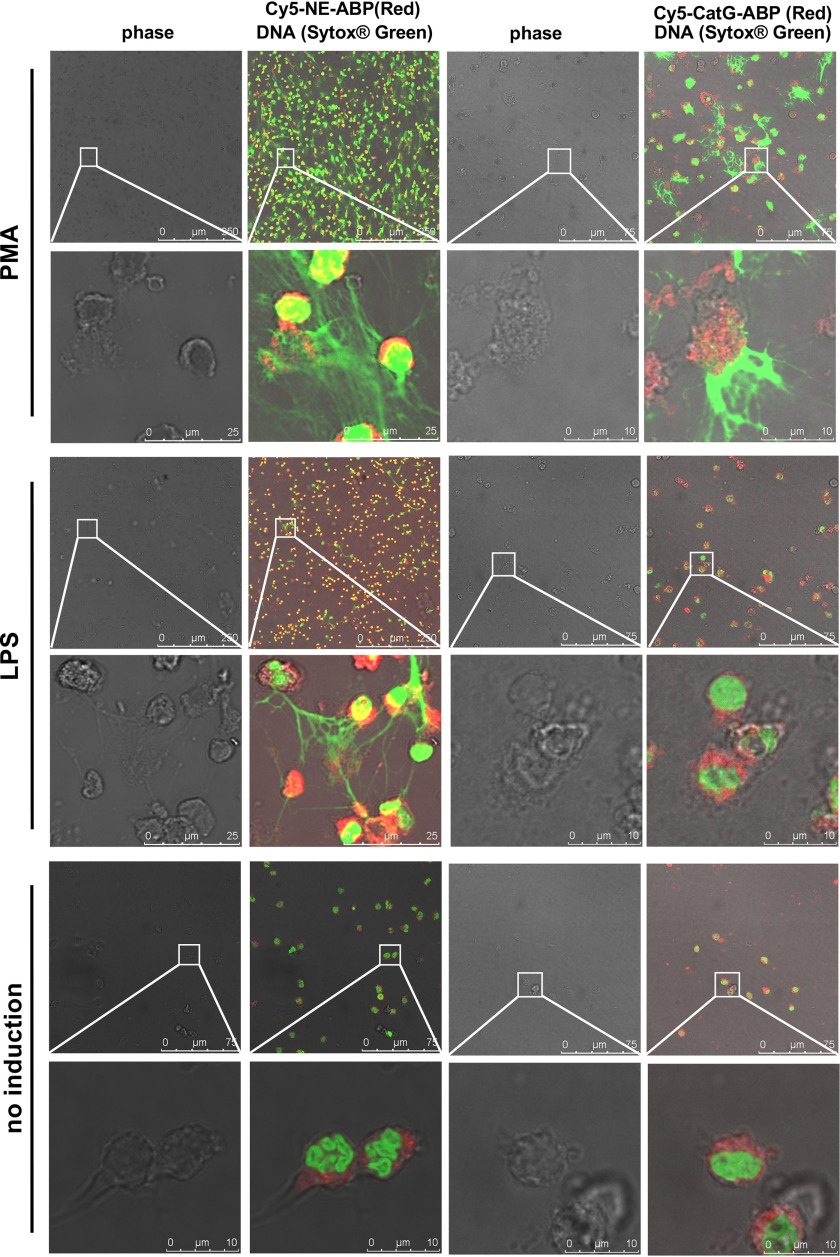
Figure 4.
Localization of active NSPs in neutrophils after induction with different stimuli. Neutrophils were treated as indicated, allowed to settle on coverslips, and incubated with Cy5-labeled specific probes (red) as indicated. Slides were fixed and DNA stained with SYTOXTM Green. Images are representative of three separate donors.

