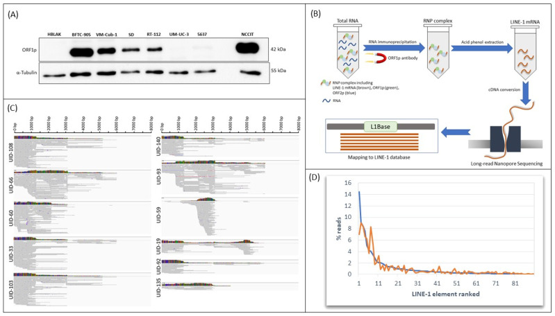
Figure 1.
Comprehensive analysis of L1 expression in VM-Cub-1 UC cells. (A) Expression of ORF1p in representative UC cell lines analyzed by Western blotting. The non-transformed urothelial cell line HBLAK was used as a negative control and NCCIT embryonal carcinoma cells were used as a positive control. α-Tubulin was used as a loading control. (B) Experimental strategy to identify expression of individual L1s in bladder cancer cells. (C) Examples of mapping results from nanopore sequencing following RIP of VM-Cub-1 cells using the highly specific antibody against ORF1p. Read alignments are shown for the elements listed in Table 1. (D) Relative expression of the 90 L1 elements detected by RIP/nanopore sequencing in the two independent experiments (blue and orange) by rank.

