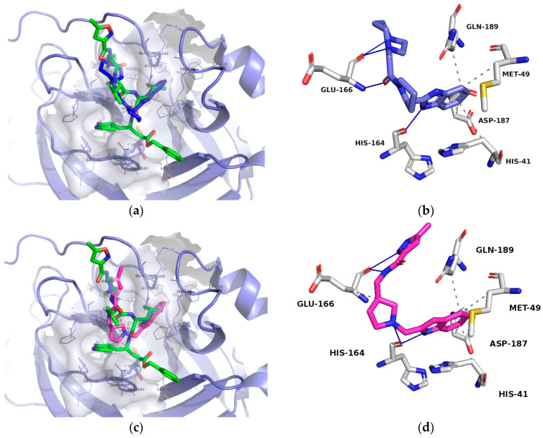
Figure 5.
(a): Compound 1 binding mode presented in blue colored stick model; (b): Compound 1 key interactions; (c): Compound 2 binding mode presented in cyan colored stick model. Reference N3 ligand from the PDB ID: 6LU7 is depicted in green-colored stick model and the protein surface around the ligand calculated. (d): Compound 2 key interactions. Hydrogen bonds are denoted as blue lines and hydrophobic interactions as grey lines.

