Figure 4.
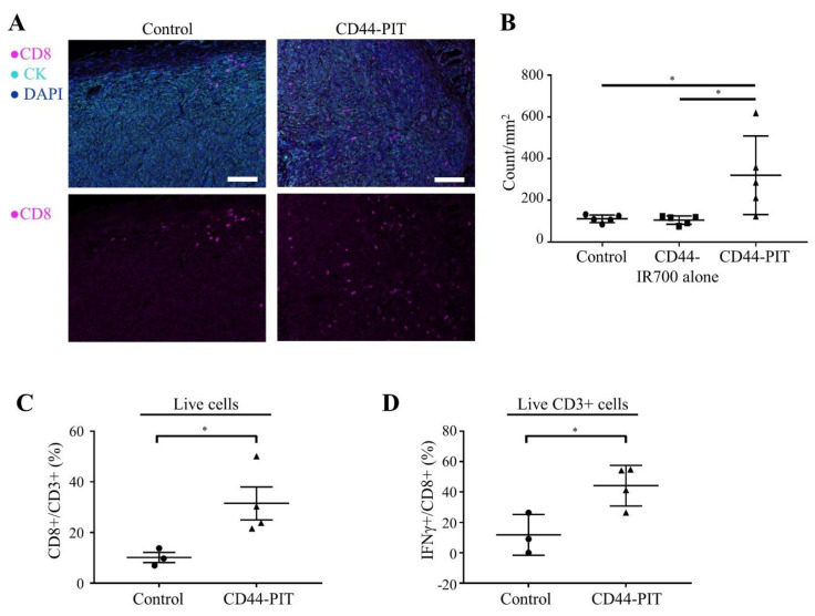
CD44-targeted NIR-PIT increased tumor infiltration of activated CD8+ T cells. (A,B) Distribution of CD8+ T cells was assessed with multiplex immunohistochemistry (IHC). (A) Representative images of multiplex IHC. Upper panels show composite images of CD8, pan-cytokeratin (CK) and DAPI staining, lower panels show single channel images of CD8 staining. CK was used to mark tumor tissue. Tumors were extracted 4 days after NIR-PIT. (×200, scale bar = 100 µm). (B) Cell number of CD8+ T cells within tumor tissue was counted in multiplex IHC images. Data are shown as cell count per mm2 (n = 5; *, p < 0.05; one-way ANOVA followed by Tukey’s test). (C) CD8+/CD3+ ratio in tumor microenvironment was examined with flow cytometry. Tumors were extracted 5 days after NIR-PIT (n = 3–4; *, p < 0.05; unpaired t-test). (D) IFN-γ+/CD8+ ratio of tumor microenvironment was examined with flow cytometry. Tumors were extracted 5 days after NIR-PIT (n = 3–4; *, p < 0.05; unpaired t-test). Control, no treatment; CD44-IR700 alone, i.v. injection of anti-CD44-IR700 only; CD44-PIT, i.v. injection of anti-CD44-IR700 with NIR light exposure. Each value represents single experiment, shown with means ± SEM.

