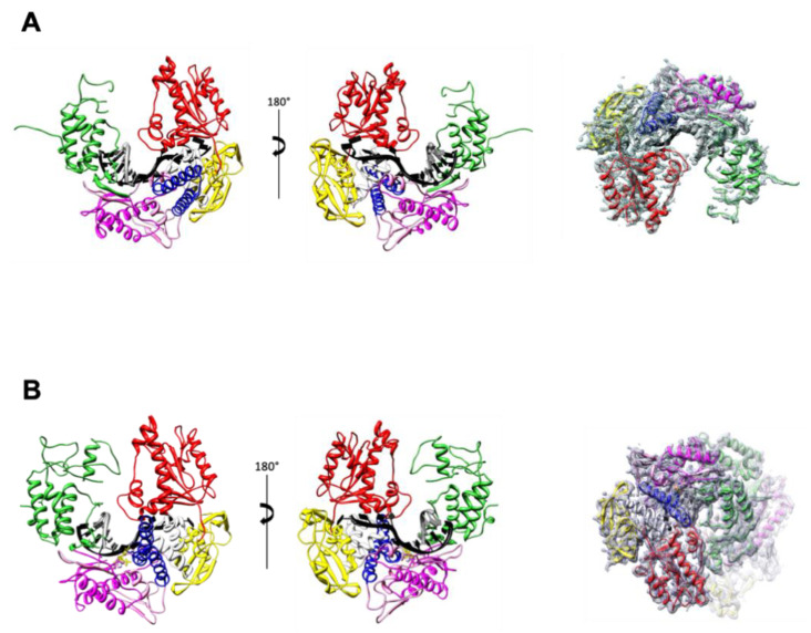
Figure 2.
TgoT_6G12 polymerase structure in binary and ternary complexes. Overall structure of TgoT_6G12 in binary (A) and ternary complex (B). On the left, front and back views of the polymerase are colored by domain. The primer and template DNA strands are colored in grey and black, respectively. The domains are labeled as follows: N-terminal domain (yellow), 3’-5’ exonuclease domain (red), palm domain (light and dark magenta represent the N-terminal and the C-terminal respectively), fingers domain (blue) and thumb domain (green). An interhelical segment between the exonuclease and the palm domain is colored in light grey. On the right, the 2Fo–Fc electron density omit map is shown and contoured at a level of 1 σ to illustrate which domains are less ordered than the others. A rotation of 180° around a horizontal axis was applied between the middle panel and the right-hand panel.

