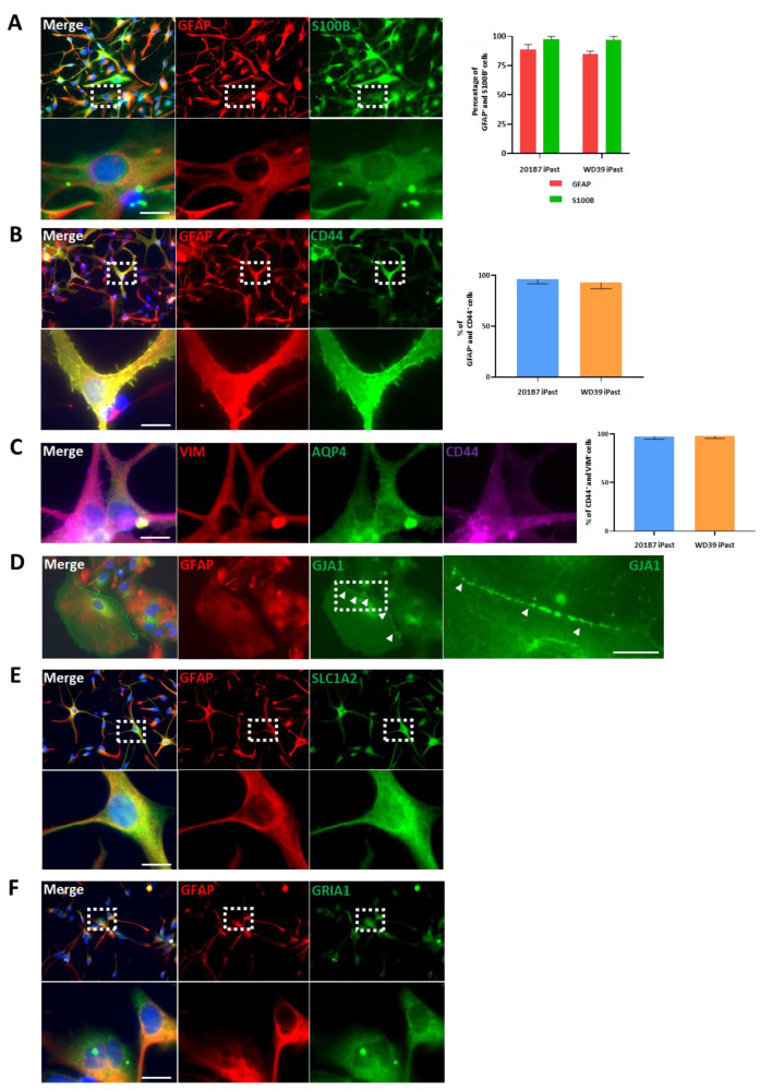
Figure 4.
Representative Immunofluorescence Stainings of 201B7 iPasts. (A) GFAP (red) and S100B (green) immunostaining of 201B7 iPasts. Lower pictures are high magnification of white dotted-line boxes. Right-hand panel, quantitative analyses of GFAP+ and S100B+ cells from 5 fields (10,000 µm²) for 201B7 and WD39 iPasts. (B) GFAP (red) and CD44 (green) immunostaining. The lower panel is a high magnification of the white dotted-line boxes. Right-hand panel, percentage of GFAP+/CD44+ from 5 fields (10,000 µm²) for 201B7 iPasts and WD39 iPasts. (C) Immunostaining for Vimentin (VIM) (red), AQP4 (green) and CD44 (magenta). Right-hand panel, analysis of CD44+/VIM+ cells from 5 fields (10,000 µm²) for 201B7 iPasts and WD39 iPasts. (D) Immunostaining for GFAP (red) and GJA1 (green). Arrowheads point to GJA1+ gap junction proteins. The picture on the extreme right is a high magnification of the adjacent white dotted-line box. (E) Immunostaining for GFAP (red) and SLC1A2 (green). The lower panel represents a high magnification of the corresponding white dotted-line boxes. (F) Immunostaining for GFAP (red) and Glutamate Ionotropic Receptor AMPA Type Subunit 1 (GRIA1) (green). Bottom pictures are high magnifications of white dotted-line boxes. Nuclei are stained with Hoechst 33258. n = 3 independent astrocytes induction experiments. Scale bars: 10 µm.

