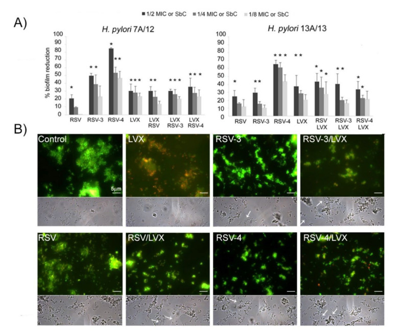
Figure 3.
Effect of RSV, its derivatives (RSV-3 and RSV-4), and LVX at sub-inhibitory concentration (1/2, 1/4, 1/8MIC) and their combinations at sub-synergistic concentrations (SbC), against H. pylori 7A/12 and H. pylori 13A/13 biofilm formation. (A) Percentage of biofilm reduction of resistant strain 7A/12 and MDR H. pylori 13A/13 after treatments. * Statistically significant values with respect to the control. (B) Representative fluorescence (after Live/Dead staining) and phase contrast light microscopy images of H. pylori 13A/13 biofilm treated with 1/4 MIC LVX, 1/4 MIC RSV, 1/4 MIC RSV-3, and 1/4 MIC RSV-4 and 1/4 sub-synergistic combinations of RSV, RSV-3, or RSV-4 and LVX and the untreated sample (control). Viable cells exhibit green fluorescence while dead cells exhibit red fluorescence. Arrows indicate the elongated forms of H. pylori cells after treatment with RSV, RSV-3, and RSV-4 alone and combined with LVX. Original magnification 1000× (scale bar: 5 μm).

