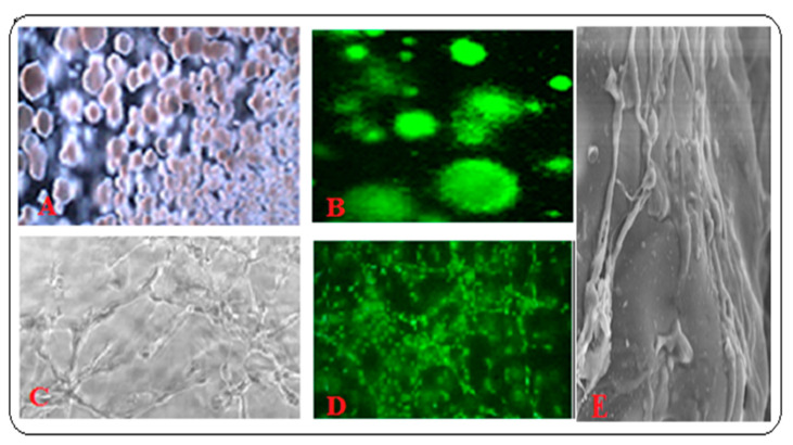
Figure 5.
HSC from BM grown on PLGA-hydroxyapatite (HA) scaffold: (A) Bright field image of the spheroid colonies; (B) Fluorescent image after staining the colonies with acridine orange-ethidium bromide stains; (C,D) 3-D BM- MSCs and HUVEC co-cultures on PLGA-HA and Matrigel scaffold, (E)- SEM image of MVs. All other microscope images were taken using 5× objective.

