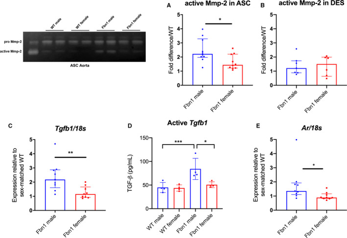
Figure 3. MMP‐2 activity, Tgfb1, and androgen receptor gene expression in Fbn1C1039G/+ and WT aortic root/ascending and descending aortas from each sex.

MMP zymography in (A) aortic root/ascending (ASC) and (B) descending (DES) aortic specimens from 16 week‐old Fbn1C1039G/+ mice (Fbn1) and littermate wild type control mice (WT) (n=10 ASC specimens per group, n=7 male and n=6 female DES specimens). Values expressed as fold difference compared with sex‐matched WT. C, RT‐PCR results for TGF‐β1 ligand (Tgfb1). D, ELISA results for active Tgfb1 ligand in 16‐week old ASC tissue. E, RT‐PCR for androgen receptor (AR) in ASC from Fbn1 at age 16 weeks (WT male=8; WT female=8; Fbn1 male=9; Fbn1 female=9). mRNA expression values are normalized to 18S as a loading control housekeeping gene and reported as fold change compared with WT using the 2−ΔΔCt method. Results presented as median±interquartile. Mann–Whitney U test used for the comparison between sex within each genotype. *P≤0.05, **P<0.01, *** P<0.001. MMP indicates matrix metalloproteinase; RT‐PCR, reverse transcription polymerase chain reaction; and TGF‐β1, transforming growth factor beta 1.
