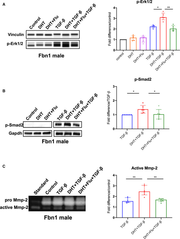
Figure 4. p‐Erk1/2, p‐Smad2 signaling and MMP‐2 activity in Fbn1C1039G/+ aortic root/ascending derived smooth muscle cells following hormone treatment.

A, Phosphorylated‐Erk1/2 (p‐Erk1/2) (n=5, each group) and (B) phosphorylated‐Smad2 (p‐Smad2) (n=6, each group) in aortic root/ascending (ASC) aorta derived SMC from Fbn1C1039G/+ (Fbn1) male mice treated with dihydrotestosterone 10 nmol/L (DHT), DHT+flutamide 1 μmol/L (Flu), TGF‐β 5 ng/mL, TGF‐β+DHT, TGF‐β+DHT+Flu or vehicle control. Relative protein level quantified using vinculin or Gapdh as loading control. Values expressed as fold difference compared with control for p‐Erk1/2. Values expressed as fold difference compared with TGF‐β for p‐Smad2 since no signal detected for control‐treated SMC. C, MMP‐2 activity in cell culture supernatants of ASC derived SMC from Fbn1 male mice treated with listed hormones or vehicle control (n=6, each group). Values expressed as fold difference compared with vehicle control or TGF‐β with representative pictures. Results presented as median±interquartile. Kruskal–Wallis test with Dunn's posttest was used for multiple comparison among the groups: control vs DHT vs DHT+Flu, TGF‐β vs TGF‐β+DHT vs TGF‐β+DHT+Flu. *P≤0.05, **P<0.01. Erk indicates extracellular‐signal‐regulated kinase; MMP, matrix metalloproteinase; and TGF‐β, transforming growth factor beta.
