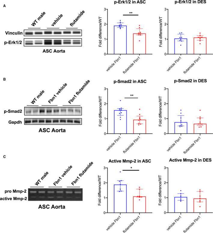
Figure 7. p‐Erk1/2, p‐Smad2 signaling and MMP‐2 activity in aortic root/ascending and descending aortas from Fbn1C1039G/+ male mice treated with flutamide.

A, Automated immunoblotting of phosphorylated‐Erk1/2 (p‐Erk1/2) (flutamide‐treated Fbn1 males=8; vehicle control‐treated Fbn1 males=8) and (B) western blotting of phosphorylated‐Smad2 (p‐Smad2) (n=9, each group) in aortic root/ascending (ASC) and descending (DES) aortic specimens from Fbn1C1039G/+ male mice treated with vehicle control or flutamide euthanized at age 16 weeks. Vinculin or Gapdh served as loading control. Values expressed as fold difference compared with littermate wild type control (WT) males. C, Gelatin zymography in ASC and DES aortic specimens from Fbn1C1039G/+ male mice treated with vehicle control and flutamide (n=7, each group) at age 16 weeks. Values expressed as fold difference compared with WT males. Results presented as median±interquartile. Mann–Whitney U test was used for the comparison between the groups *P≤0.05, **P<0.01. Erk indicates extracellular‐signal‐regulated kinase; and MMP, matrix metalloproteinase.
