
Figure 1. MicroRNAs significantly more or less expressed in the human SN in comparison with right atrial muscle.
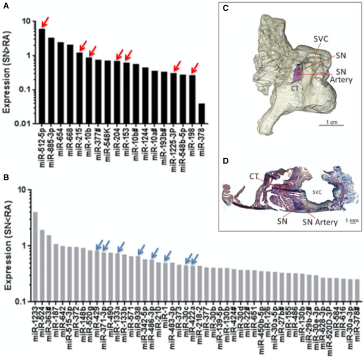
A, Expression of 18 microRNAs that are significantly more abundant in the sinus node (SN) compared with atrial muscle. B, Expression of 48 microRNAs that are significantly less abundant in the SN compared with atrial muscle (n=7; P<0.05). C, Three‐dimensional model of the human right atrium showing the location of the SN around the SN artery (similar to Stephenson et al, 2017 study). 1 The SN is stained blue by the Masson trichrome stain, because of its high content of fibrous tissue. D, Masson trichrome stained section through the SN cut perpendicular to the crista terminalis showing the location of the SN around the SN artery. See Table for key microRNAs indicated by red and blue arrows. CT indicates crista terminalis; miR, microRNA; SN, sinus node; and SVC, superior vena cava.