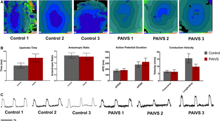
Figure 6. PAIVS hCAS exhibits insignificant electrophysiological differences from healthy control.

A, Representative isochrone maps showing the radial spread of electrical signals in healthy subject and PAIVS hCAS. B, Quantification of electrophysiological parameters of hCAS from all 6 subjects under 1‐Hz external pacing at 7 days after construction showing no significant difference in anisotropic ratio, action potential duration, and conduction velocity. n=11 for healthy controls, n=11 for PAIVS. C, Representative action potential traces for hCAS paced at 1 Hz. hCAS indicates human cardiac anisotropic sheet; and PAIVS, pulmonary atresia with intact ventricular septum
