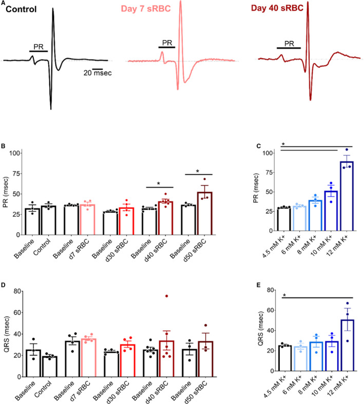
Figure 3. RBC storage age is associated with slowed atrioventricular conduction.

A, Pseudo‐ECGs recorded during sinus rhythm from isolated hearts perfused with control media (left), media supplemented with 10% sRBC collected from a day 7 unit (middle) or day 40 unit (right). PR interval time is denoted. (B,C) Atrioventricular conduction slows in the presence of day 40 and day 50 sRBC, or 10–12 mM K+. (D,E) Exposure to sRBC units had no measurable effect on ventricular depolarization time (QRS) during sinus rhythm. Mean±SEM, *P<0.05, n=3–6. RBC indicates red blood cell; and sRBC, supernatant from red blood cell units.
