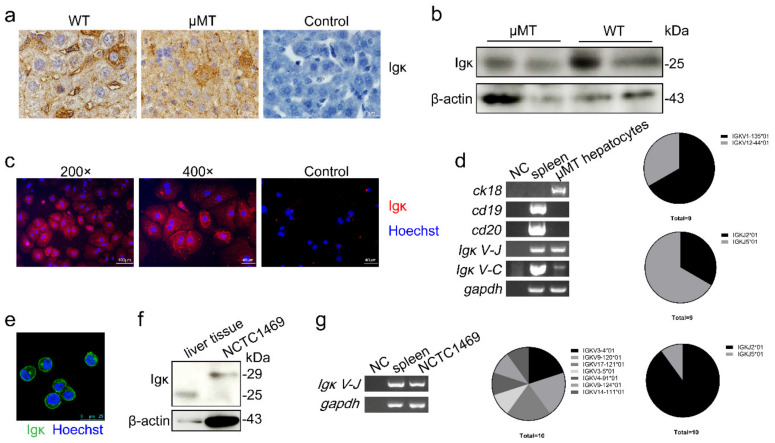
Figure 1.
Expression of Igκ in mouse primary hepatocytes and a normal hepatocyte cell line. (a) Immunohistochemistry and (b) Western blot analysis of Igκ in wild type (WT) and μMT mouse liver tissue. Control indicates isotype control. (c) The magnification and immunofluorescence analysis of Igκ in primary hepatocytes in μMT mice. Rabbit IgG as isotype control. (d) Reverse-transcription PCR analysis of ck18, cd19, cd20, Igκ V-J, Igκ J-C, and gapdh in primary hepatocytes in μMT mice. WT spleen cells were used as the positive control, and NC was used as the negative control. The frequency of Vκ and Jκ derived from primary hepatocytes in μMT mice is displayed on the right. (e) Immunofluorescence analysis of Igκ in the NCTC1469 cell line. (f) Western blot analysis of Igκ in μMT liver tissue and the NCTC1469 cell line. (g) Reverse-transcription PCR analysis and VκJκ rearrangement patterns of Igκ in the NCTC1469 cell line.

