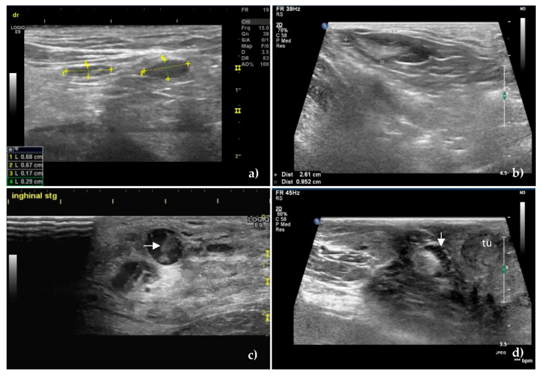
Figure 1.
B-mode ultrasound images of unaffected (a,b) and metastatic, (c,d) sentinel lymph nodes. Oval shape, S/L ratio less than 0.5, homogeneous echostructure and hyperechoic hilum of unaffected superficial inguinal sentinel lymph nodes in (a) and axillary sentinel lymph node in (b). The numbers in the image and the lower left corner of Figure 1a represent the long axis (1 and 2) and short axis (3 and 4) measurements of the examined SLNs. The distance between the two “+”signs in Figure 1b represents the measurement of the long axis, and the distance between the two “×” signs represents the measurement of the short axis of SLN. The values are found in the lower left corner of Figure 1b. (c) Metastatic superficial inguinal sentinel lymph node showing rounded shape, hypoechoic pattern, and inhomogeneous echostructure with coagulation necrosis inside of lymph node (horizontal arrow) as an echogenic structure which leaves no shadows. (d) Cortical thickening of a metastatic superficial inguinal sentinel lymph node (down arrow) located near a tumor—tu.

