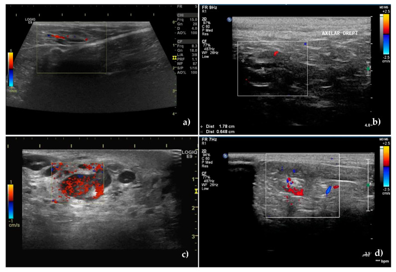
Figure 2.
Vessels location and distribution assessed by Color Doppler ultrasound. (a) Central, hilar vessels of unaffected superficial inguinal sentinel lymph node and (b) unaffected axillary sentinel lymph node. (c) Presence of neovascularisation with an abnormal, hilar, and peripheral distribution of vessels in a metastatic superficial inguinal sentinel lymph node. (d) Mixed hilar and peripheral pattern with the parenchymal subcapsular location of vessels in a metastatic axillary sentinel lymph node.

