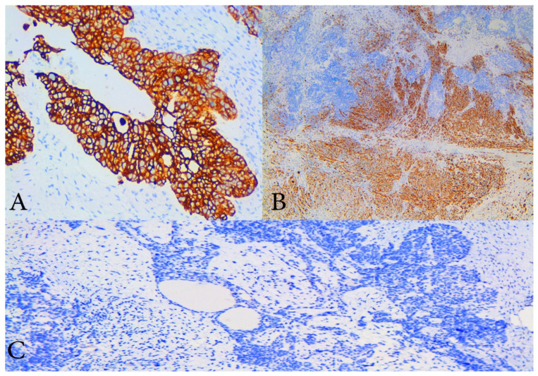
Figure 4.
Immunohistochemical staining (IHC). (A) IHC with cytokeratin 7—diffuse and strong cytoplasmic positivity in malignant epithelial cells of high- grade serous component of the tumour, ×200 magnification; (B) IHC with desmin which shows cytoplasmic positivity of sarcomatous component with smooth muscle differentiation, ×100 magnification; (C) IHC with p53 which shows that both components are negative, magnification ×200.

