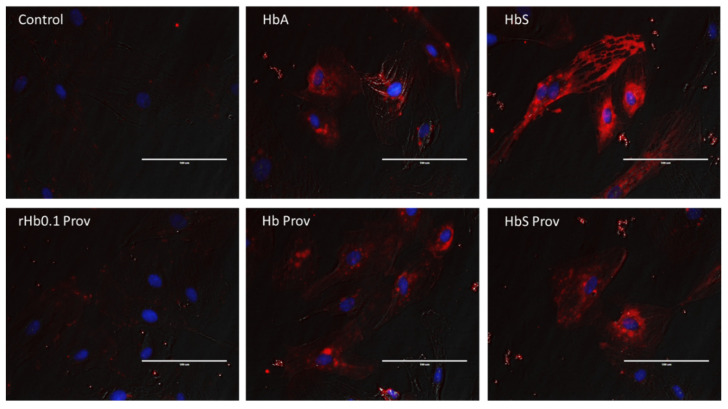
Figure 4.
Immunohistochemistry of HO-1 expression in HPAECs. HPAECs were grown on coverslips and incubated with or without HbA, Providence (βK82D), HbS, HbS Providence (βE6V/βK82D), or crosslinked Providence (rHb0.1/βK82D), at equimolar concentration (100 µM), for 12 h. Immunohistochemistry was done in paraformaldehyde fixed HPAECs, using antibody against HO-1, as described in the Methods section. Representative fluorescence microscopic images of HPAEC shows HO-1 expression (red fluorescence, AlexaFluor 595). Cell nuclei appear as blue (4′,6-diamidino-2-phenylindole, DAPI). Fluorescence images were merged onto the corresponding phase contrast views to indicate cellular structures. All images are representative of several similar fields obtained from two separate experiments. White line indicates 100 µm.

