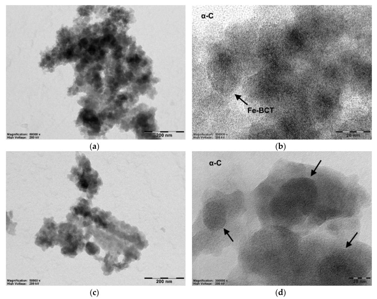
Figure 5.
(a) Transmission electron microscopy (TEM) micrograph showing an aggregate of amorphous neat Fe-BTC nanoparticles. (b) The nanoporous network of Fe-BCT at higher magnification. (c) TEM micrograph of an aggregate of neat Fe-BTC after taxol encapsulation. (d) Taxol bounded Fe-BTC nanoparticles (arrows), with their nanoporous network still visible.

