Figure 1.
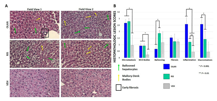
Atypical antipsychotic (AA) treatment led to histopathologic changes consistent with early non-alcoholic fatty liver disease (NAFLD). Hematoxylin and eosin staining showed several histopathologic changes at 20× magnification following AA treatment relative to vehicle (VEH)-treated controls (A). These included micro- and macrosteatosis, ballooned hepatocytes (green arrows), Mallory–Denk bodies (yellow arrows), and small areas of early fibrosis (boxed). Two representative field views are shown. Statistical significance (*, **) in lesion scores between AA-treated mice and VEH-treated mice was found for microsteatosis, Mallory–Denk body formation, and formation of inflammatory foci (B).

