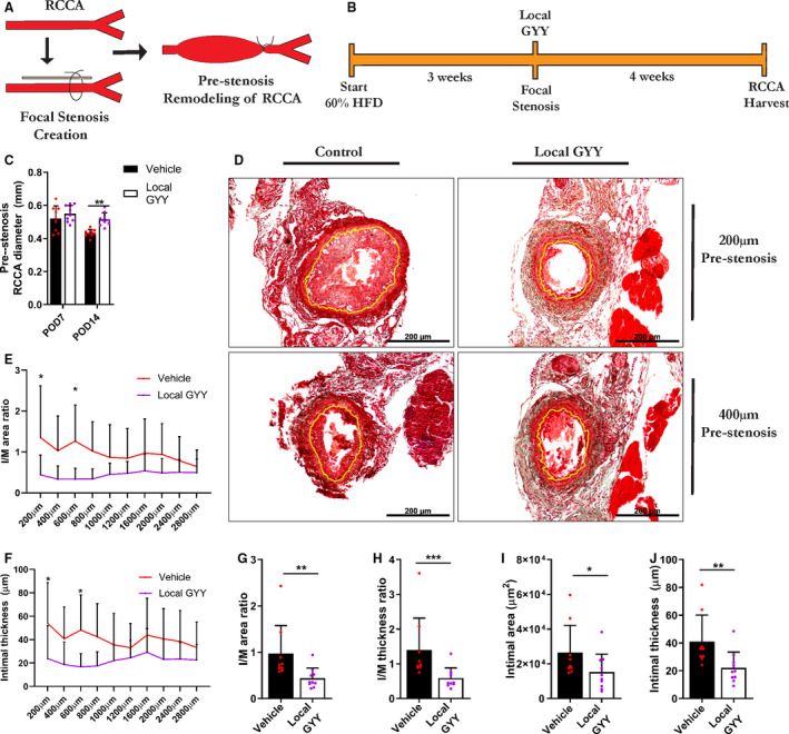
Figure 3. Local application of the H2S donor GYY mitigates injury‐induced arterial intimal hyperplasia.

A, Schematic depiction of surgical procedure, with partial ligation of the RCCA and resulting remodeling proximal from the stenosis. B, Experimental outline. C, Prestenosis diameter of RCCA at POD 7 and POD 14 in vehicle and local GYY treated animals, via 2‐way analysis of variance, n=9 to 10/group. D, Masson‐Trichome staining of RCCA cross‐sections at POD 28 after focal stenosis, with yellow lining indicating the intima‐media border. Scale bars are 200 µm as indicated. E through F, Measurement of I/M area and thickness ratio respectively at regular intervals proximal of stenosis at POD 28. G through H, Morphometric analysis of prestenosis RCCA, via Mann–Whitney test, n=9 to 10/group. G, I/M area. H, I/M thickness ratio. I, Intimal area. J, Intimal thickness. Data represented as Mean±SD. *P<0.05, **P<0.01, ***P<0.001. GYY indicates hydrogen sulfide prodrug; HFD, high‐fat diet; H2S, hydrogen sulfide; I/M, intimal/media; POD, postoperative day; and RCCA, right common carotid artery.
