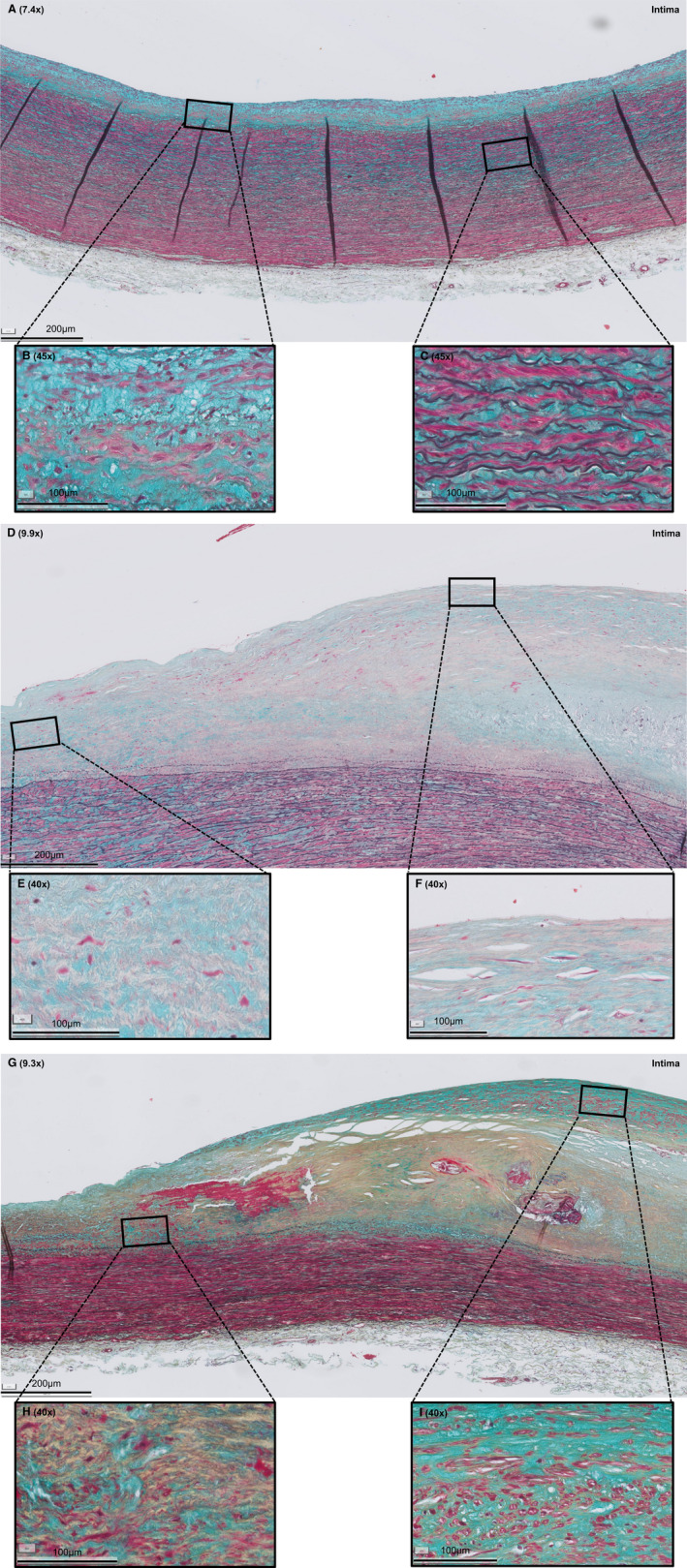
Figure 1. Histologic overview (Movat pentachrome staining) of selected representative sections of adaptive intimal thickening (AIT), late fibroatheroma (LFA), and fibrotic calcified plaque (FCP).

A, AIT is characterized by a thickening intima, consisting of smooth muscle cells (SMCs) in a proteoglycan‐rich matrix (B) and a normal media and adventitia (C). D, LFA is characterized by a necrotic core of cellular debris and cholesterol crystals that is covered by a multilayered fibrous cap, consisting of SMCs in a collagenous proteoglycan‐rich matrix with infiltration of inflammatory cells (F). E, Shoulder regions. G, FCP is characterized by extensive fibrosis, a condensated (former) necrotic core, and ample calcification (H) and neointimal formation (I). Legend to the Movat staining: red, SMCs/fibrin; violet, leukocytes; black, elastin; blue, proteoglycans/mucins; yellow, collagen. Various shades of green reflect colocalization of collagen (yellow) and proteoglycans (blue).
