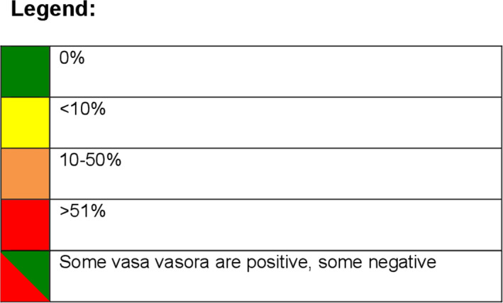
Figure 4. Semiquantitative evaluation of single immunohistochemistry (IHC) stainings.

The presence of immunohistochemical markers is appreciated in 6 zones of the vessel wall: intima (I), inner media underlying lesion (M1), middle media (M2), outer media (M3), adventitia (Ad), thin‐walled, venous‐type vasa vasorum (VV thin), and thick‐walled artery‐type vasa vasorum (VV thick). In late fibroatheroma (LFA), the intima is divided in a central cap region (Cap) and shoulder regions (Sh); and in fibrotic calcified plaque (FCP), the neointima (Neo) overlaying the fibrous cap is considered. AIT indicates adaptive intimal thickening; FAP, fibroblast activation protein; FSP, fibroblast‐specific protein; α‐SMA, α‐smooth muscle actin; SM22α, smooth muscle protein 22α; Smemb, nonmuscle myosin heavy chain; SM‐MHC, smooth muscle myosin heavy chain; and Thy1, thymocyte differentiation antigen 1.
