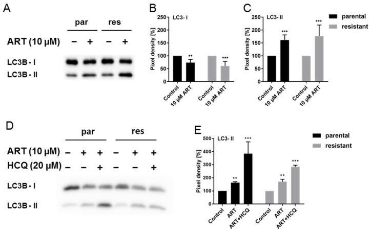
Figure 7.
Autophagy: Expression of LC3B-I and LC3B-II in parental (par) and cisplatin-resistant (res) T24 cells after 24 h exposure to 10 µM ART alone (A) or in combination with 20 µM HCQ (D). A representative Western blot of n = 4 is shown. Each protein analysis was normalized by a total protein staining control. Pixel density analysis of the protein expression (B,C,E), compared to the untreated controls (set to 100%) is illustrated. Error bars indicate standard deviation (SD). Significant difference to untreated control: ** p ≤ 0.01, *** p ≤ 0.001. n = 4. For detailed information regarding the Western blots see Figure S3.

