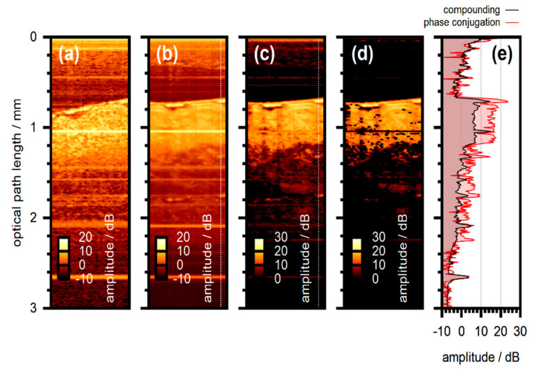
Figure 13.
Wavefront shaping at layered scattering phantom. Phase conjugation with biological tissue. (a) Conventional OCT scan. (b) Image captured with a speckle-compounding algorithm to reduce speckle contrast. (c) Scan acquired with phase conjugation. Number of modes N = 256. (d) Phase conjugation with additional artefact suppression. (e) Amplitude of the two A-scans marked by the dashed line in panels (b) and (c), respectively. The amplitude color-scale corresponds to the same dynamic range of 30 dB for all scans. Adapted from [8,106], with permission.

