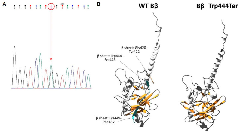
Figure 4.
(A): Electropherogram of the heterozygous mutation in exon 8 of the FGB gene (c.1421G>A), (B): Analysis of the Bβ Trp444Ter mutant model. Differences between the secondary structure of the WT Bβ chain and the Bβ Trp444Ter are indicated. The amino acids missing in the mutant chain are displayed in blue. Alpha helices and beta sheets are colored in grey and orange, respectively, while the remaining residues are assigned as dark grey. Amino acids are numbered according to the mature protein sequence. Images were prepared using Swiss-PdbViewer 4.1.0, POV-Ray, and PDB file 1FZA [9].

