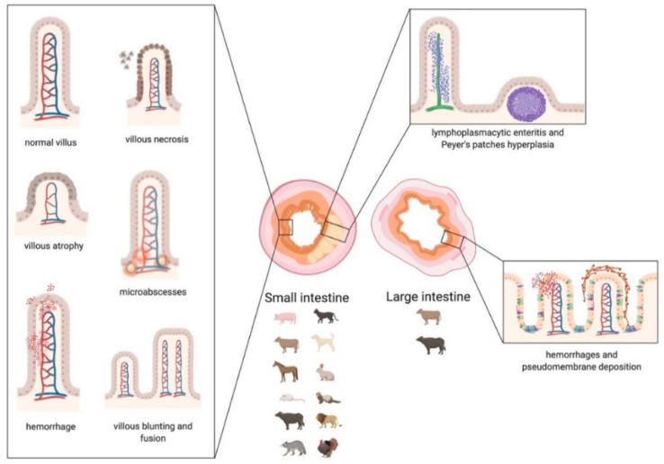
Scheme 4.
Graphic representation of the main lesions detected in small and large intestine associated with Coronavirus infections in animals. Small intestine: in the left box, the morphologic changes of the villi are represented. In the upper right box, the distribution of the infiltrate, affecting the lamina propria and the submucosa, is represented. Large intestine: in the lower right box, the lesions affecting the mucosa of the colon are represented. The main categories of affected species that suffer damage mainly to the small or large intestine are indicated below the corresponding image. The scheme has been created with BioRender.com.

