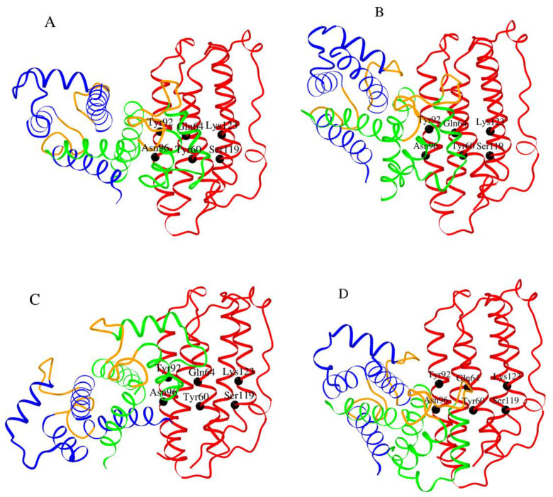
Figure 9.
The models of tertiary structures of Ca2+-loaded human S100P (panel A), S100A1 (B), S100A4 (C) and S100A6 (D) dimers (the monomers are shown in blue/green) bound to a molecule of human IFN-β (red) built using GRAMM-X protein–protein docking software v.1.2.0 [58]. The Ca2+-binding loops are shown in orange. The contact residues of IFN-β are labelled.

