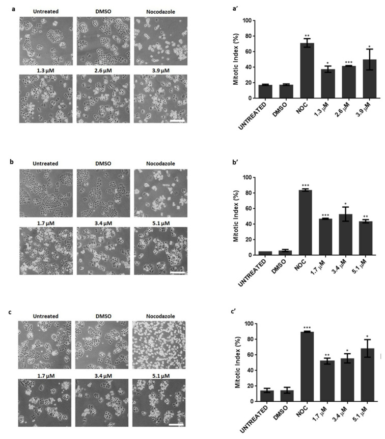
Figure 2.
Cancer cells arrested in mitosis, in response to pyranoxanthone 2 (PX2) treatment. Left-Representative phase contrast microscopy images, after 16 h treatment with PX2, at indicated concentrations, displaying accumulation of mitotic cells (rounded) in (a) A375-C5, (b) MCF-7 and (c) NCI-H460 cancer cell lines. Nocodazole was used as a positive control. DMSO was used as a compound solvent control. Bar, 10 µm. Right-Mitotic index graphs showing accumulation of mitotic cells in (a’) A375-C5, (b’) MCF-7 and (c’) NCI-H460 cancer cells with statistical relevance of * p < 0.05, ** p < 0.01 and *** p < 0.001 by unpaired t-test.

