Figure 1.
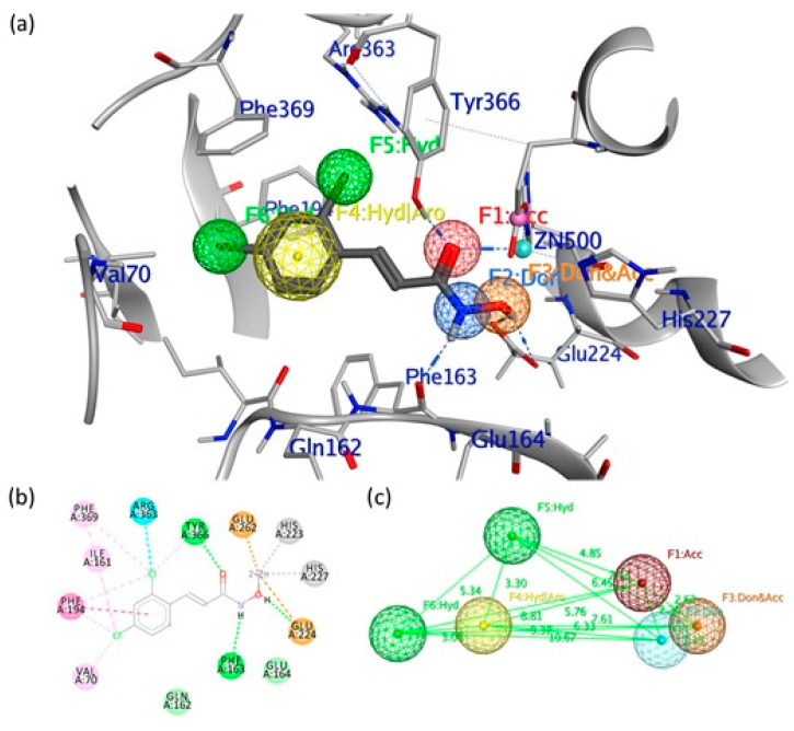
(a) Pharmacophore model generated by the Pharmit server, including hydrogen bond donors (Don) (blue/orange spheres), negatively charged oxygen atom to represent a hydrogen bond acceptor (Acc) (red sphere), the hydrophobic center (Hyd) (green sphere) and the aromatic center (yellow sphere). (b) Binding site interactions between the light chain of botulinum neurotoxin type A (LC/A) binding pocket and the co-crystallized (2E)-3-(2,4-dichlorophenyl)-N-hydroxyacrylamide (DCNHA). (c) 3D spatial distribution of the six pharmacophore features.

