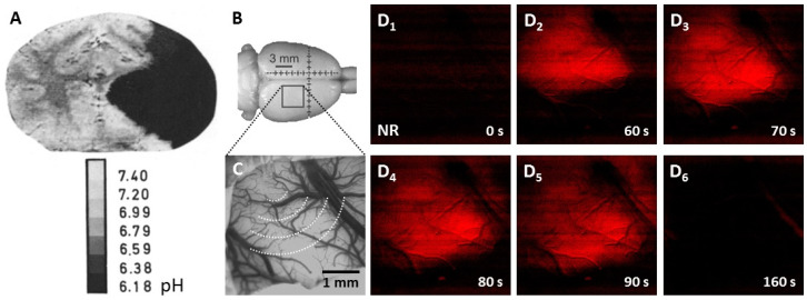
Figure 4.
Experimental tissue pH imaging in models of cerebral ischemia and spreading depolarization. (A), Umbilliferone fluorescence pH imaging 2 h after experimental middle cerebral artery occlusion in the cat. Reprinted from Csiba et al., 1983 [80], with permission from Elsevier. (B–D), Neutral red fluorescence imaging of spreading depolarization (SD) through a closed cranial window preparation over the parietal cortex of an anesthetized rat. (B), The position of the closed cranial window. (C), An intrinsic optical signal image of the exposed cortical surface at green light illumination. The schematic radial hemi-circles indicate the wave of SD. (D), Transient tissue acidosis propagating with SD, depicted by the increasing intensity of the Neutral red fluorescent signal (red) in background-subtracted, contrasted and pseudo-colored images. Time with respect to SD initiation is shown in the lower right corner of the images.

