Figure 3.
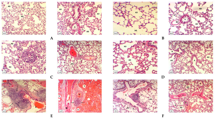
(A) Cross section of lung of normal healthy albino rats represents: (a.1) normal architecture of lung tissue, alveolar ducts (AD), sacs, alveoli (A) and capillary (arrow); (a.2) it shows normal bronchioles (B), alveolar ducts (AD), sacs and normal blood vessels (BV). (B) Cross section of lung of rats which fed on propolis extract: (b.1) it shows alveolar ducts (AD) and alveoli (A); (b.2) it illustrates normal bronchioles (B) with lumen is empty, alveolar ducts (AD), sacs and normal blood vessels (BV). (C) Cross section of another lung of low dose of aluminum silicate (5 mg/kg) induced rats: (c.1) it shows numerous lymphocytes (arrows) and alveolar ducts (AD) are noticed; (c.2) it shows congestion of blood vessels (C), numerous lymphocytes (arrows), alveolar ducts (AD) and alveoli (A). (D) Cross section of another lung of low dose of aluminum silicate (5 mg/kg) and propolis treated rats show clear signs repair of architecture of lung tissue: (d.1) it represents the alveolar ducts (AD) and blood vessels (BV) with restoration of normal lung histological architecture; (d.2) it shows alveoli (A) with more defined, mild degeneration of the bronchioles (B) and some congestion of blood vessels (C) are founded. (E) Cross section of another lungof high dose of aluminum silicate (20 mg/kg) induced rats: (e.1) it illustrates a significant accumulation of macrophages and small increases of neutrophils (arrow), formation of eosinophilic hyaline casts inside the lumen of bronchioles (B) and congestion of blood vessels (C); (e.2) it displays formation of eosinophilic hyaline casts inside the lumen of bronchioles (B), numerous lymphocytes (arrows) with fibroblastic proliferation replacing lung tissue (thick arrow) and many of vacuoles are visible. (F) Cross section of lung of high dose of aluminum silicate (20 mg/kg) and propolis treated rats: (f.1) it displays restoration of the bronchioles (B), alveolar ducts (AD), alveoli (A) are mild repair and some congestion of blood vessels (C) are stilled found; (f.2) it illustrates clear signs repair of architecture of lung tissue. The bronchioles (B), alveolar ducts (AD), alveoli (A) are mild repair, some congestion and hemorrhage of blood vessels (C) are seen. All cross sections of rat lung tissues stained with H&E (magnification power was 100 X using light microscopy).

