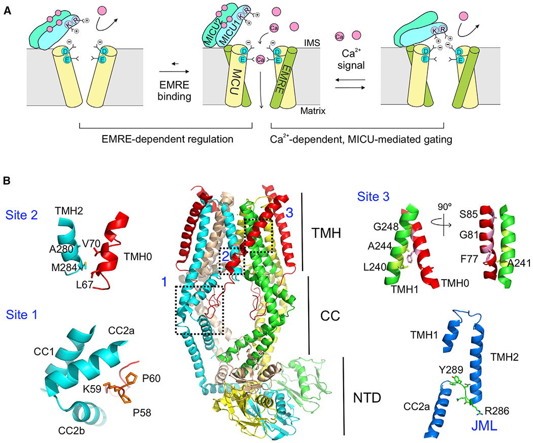
Figure 1. Uniporter Architecture and Regulation.

(A) A cartoon illustrating EMRE- and MICU-dependent regulation of the uniporter. Only 2 copies of MCU and EMRE in the MCU-EMRE tetramer are illustrated to show the Ca2+ pathway.
(B) Structure of the human MCU-EMRE subcomplex. The 3 MCU-EMRE contact sites, the JML (green, bottom right), and the JML surrounding areas (blue, bottom right) are highlighted. The second coiled-coil (CC2) in MCU is broken into halves, CC2a and CC2b. The MCU subunit in cyan shows a large fenestration between its 2 TM helices.
CC, coiled-coil; NTD, N-terminal domain; TMH, transmembrane helices.
See also Figure S1.
