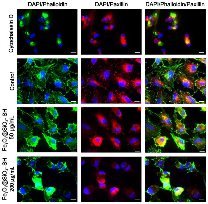
Figure 9.
Fluorescent microscopic images of A549 cells stained with Alexa Fluor 488 phalloidin (F-actin, green) and anti-paxillin (red), and counterstained with DAPI (nuclei, blue). The cells were sham-treated with H2O (negative control) or treated Fe3O4@SiO2-SH nanoparticles at 50 and 200 µg/mL. Cells treated with 0.5 µg/mL of cytochalasin D, a member of the cytochalasin fungal alkaloids that acts as a potent inhibitor of actin polymerization, were used as a reference in this assay. Experiments were performed in triplicates by using epifluorescence microscopy. Photographs from representative chambers are shown. The scale bar in the images represents 10 μm.

