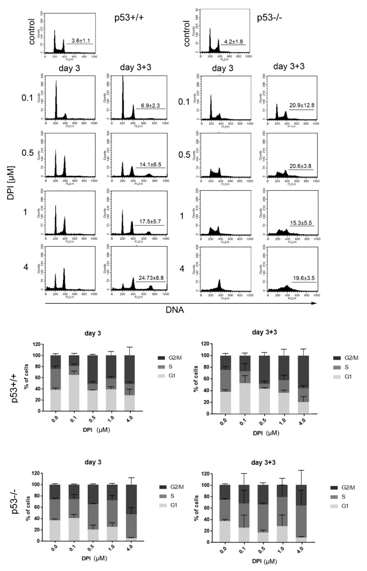
Figure 2.
DPI disturbs cell cycle progression in HCT116 p53+/+ and p53−/− cells. HCT116 cells were treated with different concentrations of DPI and cell cycle distribution was analyzed 3 days after treatment and after subsequent culture in DPI-free medium. Representative histograms (upper panel) and graphs presenting distribution of cells in different phases of cell cycle. Mean values from 4 independent experiments ±SD. The mean number (±SD) of polyploid cells is marked on each histogram.

