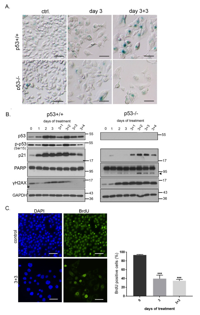
Figure 5.
DPI treatment induces senescence of HCT116 cells. (A) HCT116 cells were treated with 0.5 µM DPI for 3 days and SA-β-gal activity was analyzed; (B) the level of protein markers of senescence—p53, p-p53 and p21, and apoptosis—PARP cleavage (indicated by arrowhead) and γH2AX—was estimated at different time points after DPI (0.5 µM) treatment. (C) BrdU incorporation analysis was performed in HCT116 p53+/+ cells. The percentage of cells that were able to incorporate BrdU within 24 h of culture is presented on the graph (mean ± SD, four experiments); *** p ≤ 0.001.

