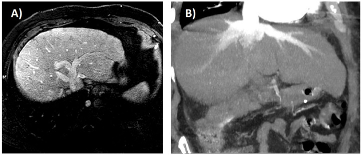
Figure 3.
(A) Idiopathic membranous inferior vena cava obstruction in a 44-year-old man. Magnetic resonance imaging shows a mildly nodular liver with altered parenchymal perfusion and dilatation of hepatic veins. (B) Severe tricuspid regurgitation in a 49-year-old man. Computed tomography scan shows dilatation of hepatic veins and reflux of contrast into the inferior vena cava and hepatic veins.

