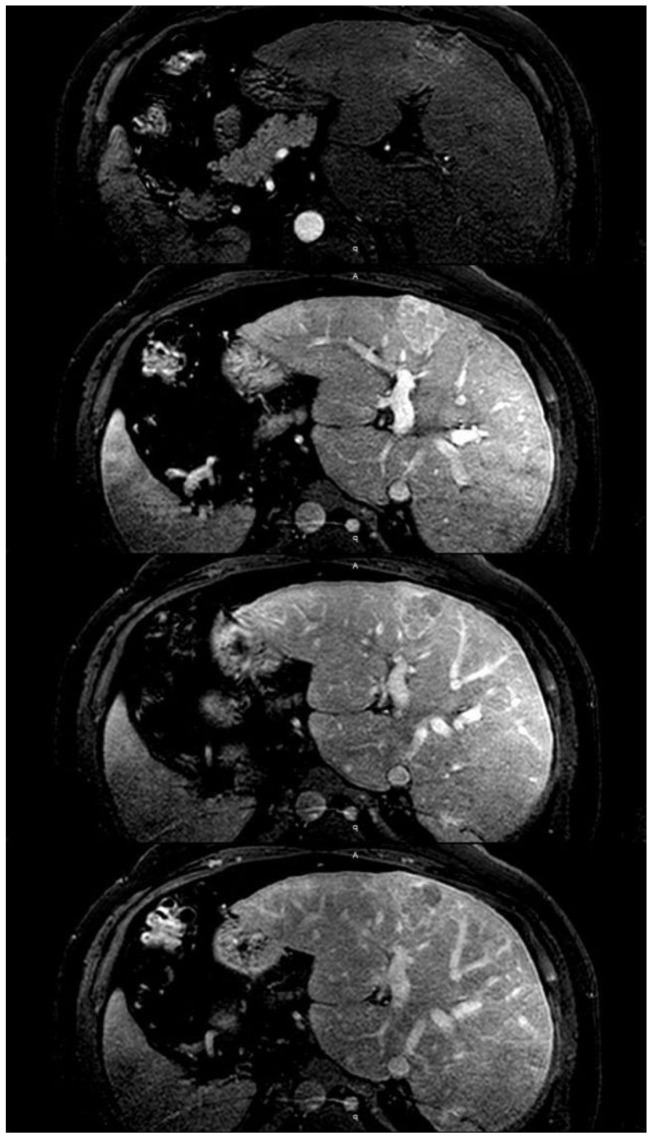
Figure 4.
Idiopathic membranous inferior vena cava obstruction in a 44-year-old man. The image shows the dynamic phase of MRI. In addition to the significant hypertrophy of segment I, magnetic resonance imaging shows a mass (3.8 cm × 4.2 cm) that after administration of intravenous contrast presents a heterogeneous enhancement in the arterial phase with washout in the portal phase. Liver biopsy showed histological changes compatible with focal nodular hyperplasia.

