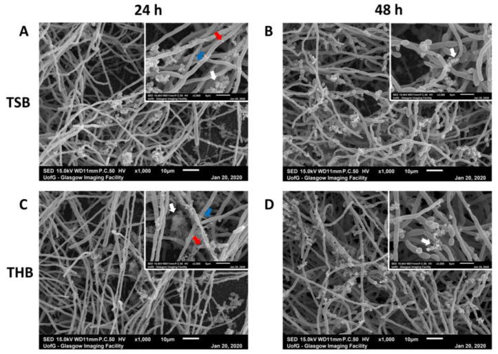
Figure 3.
Scanning electron microscopy images of multi-species biofilms. Mixed biofilms were grown in 1:1 1640/TSB (A,B) or in 1:1 RPMI/THB (C,D) + 10% FBS for 24 h (A,C) and 48 h (B,D), before being processing for scanning electron microscopy (SEM) imaging. SEM images show rich hyphal C. albicans growth with clusters of bacteria adhering the hyphae. These clusters of bacteria predominantly appear characteristic of morphological streptococci (white arrows). Blue and red arrows indicate F. nucleatum and P. gingivalis shaped colonies attached to the Candida hyphal network. Scale bar represents 10 µm and 5 µm at ×1000 and ×3500 magnification, respectively.

