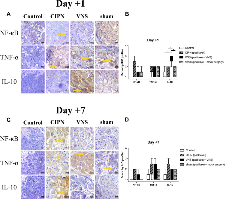
Figure 4.
Pro- and anti-inflammatory regulators in dorsal root ganglia. Immunohistochemistry was performed on dorsal root ganglia tissue taken from CIPN rats treated with vagus nerve stimulation (VNS) or not. (A, C) Representative images of staining against NF-κB, TNF-α, and IL-10 on day +1 or +7. (B, D) Immunohistochemistry score was calculated using the IHC Profiler plugin in ImageJ (see Methods). Magnification, 20X. n = 5 per group per time point. Yellow arrows indicate areas of high expression. * p<0.05.

