
Fig 4:
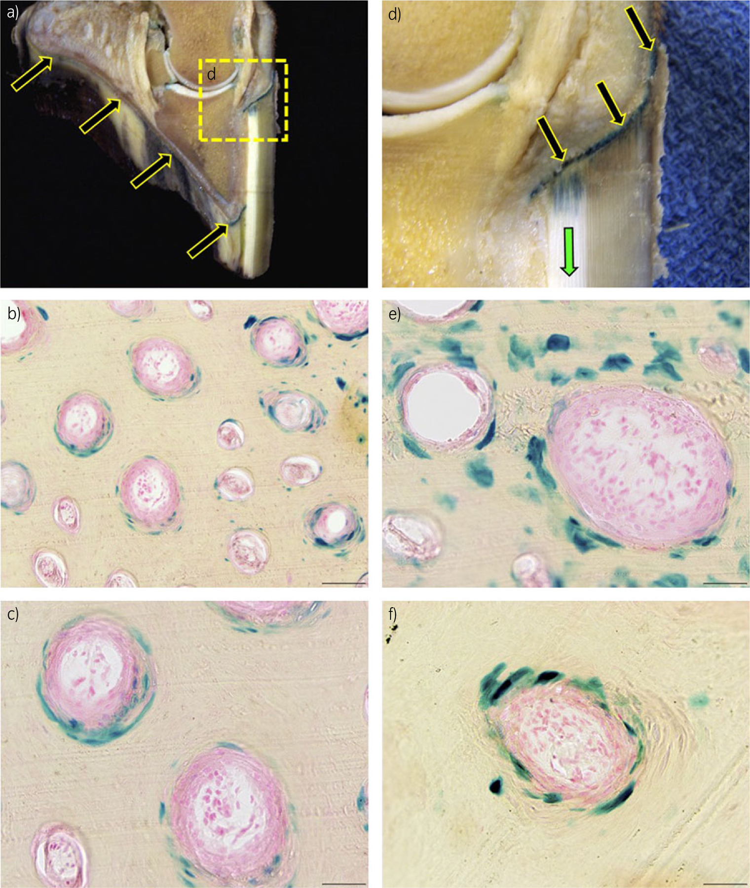
β-galactosidase (β-gal) staining, 7 days post injection. a) Sagittal section of an equine digit. β-galactosidase protein is expressed in the coronary and sole epidermal lamellae and stratum medium (arrows). Images (b), (c), (e) and (f) are representative frozen histological cross-sections of the hoof stratum medium, which display blue, nuclear β-gal staining in cells of the hoof-wall tubules. b) 9200, scale bar = 100 µm. c), e) and f) 9400, scale bar = 50 µm. d) Higher magnification of the coronary epidermis expressing β-gal.