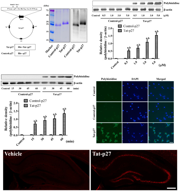
Figure 1.
Construction and delivery of Tat-p27 and Control-p27 into HT22 cells. Vectors of Tat-p27 and Control-p27 were constructed and their expressions were confirmed by Coomassie brilliant blue staining and western blot analysis for polyhistidine. Intracellular delivery of Tat-p27 and Control-p27 was confirmed by western blot for polyhistidine using various concentrations (0.5–5.0 μM) and incubation durations (15–60 min) in HT22 cells. Western blot assays were performed in at least triplicate and bar graph represents the mean ± standard deviation. Density of polyhistidine bands was analyzed using two-way analysis of variance (ANOVA) followed by a Bonferroni’s post hoc test (a p < 0.05, significantly different from the control group; b p < 0.05, significantly different from the concentration- or time-matched Control-p27 group). Localizations of intracellular-delivered Tat-p27 and Control-p27 were detected by polyhistidine immunocytochemistry in HT22 cells. Scale bar = 20 μm. Visualization of Tat-p27 and Control-p27 delivery into the hippocampus is observed 8 h after protein treatment by polyhistidine immunofluorescent staining. Scale bar = 400 μm.

