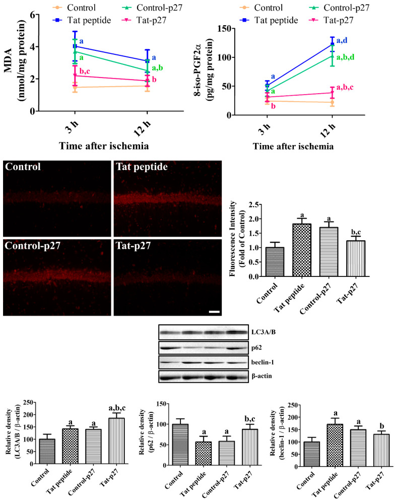
Figure 4.
Ameliorative effects of Tat-p27 and Control-p27 on ischemia-induced oxidative stress and autophagy. Reactive oxygen species (ROS)-induced malondialdehyde (MDA) and 8-iso-prostaglandin F2α (8-iso-PGF2α) levels were measured using enzyme-linked immunosorbent assay (ELISA) assay kits 3 and 12 h after ischemia in the Tat peptide, Control-p27, and Tat-p27 groups. ROS were visualized with dihydroethidium (DHE) fluorescence in the hippocampal CA1 region 3 and 12 h after ischemia in these groups. Scale bar = 50 μm. Autophagy was evaluated using western blot for microtubule-associated 1A/1B light chain 3A/B (LC3A/B), beclin-1, and p62 12 h after ischemia. Data were analyzed using one-way or two-way ANOVA followed by a Bonferroni’s post hoc test (n = 5 per group; a p < 0.05, significantly different from the control group; b p < 0.05, significantly different from the Tat peptide group; c p < 0.05, significantly different from the Control-p27 group; d p < 0.05, significantly different from 3 h post-ischemic group). Bar graph represents the mean ± standard deviation.

