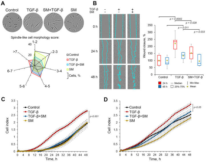
Figure 5.
SM effectively inhibits TGF-β-stimulated EMT-associated processes in A549 cells. (A) SM increases the percentage of roundish-shaped cells undergoing TGF-β-induced EMT. A549 cells were incubated with the presence of TGF-β and/or SM for 48 h followed by the evaluation of cellular morphology by phase contrast microscopy and the calculation of spindle-like cell morphology score, according to the formula (1), using ImageJ tool. (B) SM effectively decreases wound healing rate of TGF-β-stimulated A549 cells. The scratched cell monolayers were incubated with the presence or absence of TGF-β (50 ng/mL) and SM (0.5 µM) for 24 h and 48 h after that the analysis of wound closure capacity was carried out using phase contrast microscopy data. (C) SM inhibits TGF-β-stimulated motility of A549 cells. The cells were seeded in the upper chamber of cell invasion/migration (CIM)-Plate and treated with TGF-β (50 ng/mL) and/or SM (0.5 µM) for 48 h. Fetal bovine serum (FBS)-mediated migration of the cells to the lower chamber was analyzed using the xCELLigence Real-Time Cell Analyzer Dual Plates (RTCA DP) system. (D) SM inhibits TGF-β-stimulated invasion of A549 cells. The cells were seeded in the upper chamber of CIM-Plate, the bottom of which was covered by Matrigel, and treated with TGF-β (50 ng/mL) and/or SM (0.5 µM) for 48 h. Invasion of the cells to lower chamber, containing 10% FBS, was analyzed by xCELLigence RTCA DP instrument.

