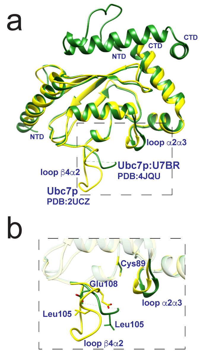
Figure 6.
Structural representations (a) of Ubc7p (yellow, PDB 2UCZ) overlaid with Ubc7p:U7BR (green, PDB 4JQU) highlighting the minimal differences within the two structures with a dashed box. A zoomed-in representation (b) showing loop shifts away from the Cys89 once U7BR binds to Ubc7p. This interaction also introduces changes in loop , highlighted by the structural differences in side-chains Leu105 and Glu108. Figure adapted from Metzger et al. [84].

